Antibodies (Ab) are large, globulin proteins produced by B cells and plasma cells as a part of the adaptive immune response. They are Y-shaped in structure, with the two antigen-binding fragments (Fab) forming the arms and the central constant fragment (Fc) joining them. At the end of each Fab is the variable region and, at the tip, is the complementary-determining region (CDR), which is the binding site for the epitope on the target antigen (Ag).
Uses of mAbs
Monoclonal antibodies (mAb) are highly selective therapies with applications in multiple medical fields. All Abs in an mAb product contain identical CDRs and thus target a single epitope on a single Ag with high specificity. The target Ag determines the application.
mAbs developed against microbial Ags can be used to treat or prevent infection by directing the adaptive immune response against the known pathogen. mAbs targeting endogenous cytokines or receptors can be used to modulate the immune system in immune-mediated diseases by interfering with ligand binding, downregulating receptors, and/or depleting target immune cells.
In human oncology, mAbs have been used extensively through several different approaches. The two most common are:
- targeting cell-surface markers overexpressed by certain cancers and inducing
cytotoxicity and; - interfering with tumor-induced immunosuppression in the tumor microenvironment
through blockade of immune checkpoints.
Development and Pharmacology of mAbs
Developed in the 1970s, the original technique for creating mAbs was the hybridoma method. This involved immunizing a mouse against the target Ag and hybridizing harvesting B cells with multiple myeloma cells to create an immortalized cell line (a “hybridoma”) that consistently produced the desired mAb. These original mAbs were completely murine in structure and so carried a high risk of hypersensitivity reaction, particularly upon repeat administration.
Subsequent molecular techniques were developed to replace the immunogenic portions of the protein with amino acid sequences of the patients’ species. Chimera mAbs replace the Fc and part of the Fab sections with self-protein; and humanized (caninized/felinized) mAbs replace all but the CDR. In people, fully human mAbs have been developed using transgenic rodents, but this technique has not been applied in any commercial veterinary mAb to date.
As protein-based therapies, mAbs have some unique pharmacokinetic properties. They are large macromolecules and susceptible to proteolytic degradation; thus, they are not orally bioavailable and must be administered parenterally. When administered IM or SC, mAbs are absorbed by the lymphatics and enter circulation; this process takes time, so maximum concentrations and effect may not be seen until a few days after administration.
The volume of distribution is low, indicating the mAbs primarily reside in the vasculature and reach the target tissue by extravasation. Immunoglobulins are eliminated by proteolysis in endothelial lysosomes; however, IgG molecules are more resistant to degradation because they bind to the FcRn receptor on the inner surface of endosomes and are trafficked back to the cell surface and re-released. Therapeutic mAbs are derived from IgG subtypes and so have prolonged half-lives due to this mechanism. These long half-lives allow for prolonged dosing intervals.
mAbs are generally very safe. However, they are protein molecules and so can be immunogenic. With the advent of chimeric and caninized/felinized mAbs, hypersensitivity reactions are rare, but the patient’s immune system can still produce antibodies against the therapeutic protein. These anti-drug antibodies have the potential to decrease efficacy by binding the therapeutic mAb and removing it from circulation.
As proteins, mAbs are also at increased risk for degradation, so storage and administration instructions should be followed carefully. In the current drug nomenclature, mAb product names end with the suffix “-mab” and those intended for veterinary use end in “-vetmab.”
Cytopoint
Released in 2017, Cytopoint (lokivetmab) was the first of the modern veterinary mAb therapies on the market. Cytopoint is a caninized mAb that targets and inhibits canine IL-31, a pruritogenic cytokine released primarily by type 2, T helper cells as a part of the inflammatory response in atopic dermatitis. IL-31 binds its receptors on peripheral sensory neurons and the dorsal horn of the spinal cord, transmitting the “itch signal” to the central nervous system. This causes the animal to scratch, which exacerbates the local inflammation and causes a vicious cycle of itching.
By neutralizing IL-31, Cytopoint prevents it from binding the receptor and interrupts this pathway. Interestingly, IL-31 receptors are expressed on other cell types, including immune cells and epithelial cells, and in other body tissues, such as the respiratory and gastrointestinal tracts. Thus, it is likely that IL-31 has functions beyond the neuroimmune axis, and Cytopoint might be useful for treating certain other inflammatory conditions. However, this requires further study.
Cytopoint has demonstrated efficacy in the treatment of canine atopic dermatitis. In a placebo-controlled trial, a single Cytopoint injection led to an average 57% reduction in owner-assessed itchiness in atopic dogs compared to 21% in saline control. The effect was detectable within 1 day of the injection, reached maximal effectiveness by day 7, and persisted for at least 28 days.
Monthly Cytopoint injections elicited similar pruritus control compared to oral cyclosporine (5 mg/kg/d) and extended dosing (>4 to 6 months) resulted in remission of atopic dermatitis in greater than 50% of dogs.
It can also be used in other forms of allergic dermatitis, such as pododermatitis, otitis, and flea allergy dermatitis. Pruritus reduction and duration of action are dose-dependent, so it is recommended to administer Cytopoint at a minimum dose of 2 mg/kg. Cytopoint is generally administered monthly; however, it is possible to extend the dosing interval to 6 or 8 weeks in some dogs. Safety trials have not documented specific adverse effects, but hypersensitivity reactions could occur.
Solensia
Solensia (frunevetmab) is a felinized mAb that targets nerve growth factor (NGF) and is approved for the management of osteoarthritis (OA) in cats. NGF is an important modulator of nociception in chronic pain states and is released by damaged peripheral tissues. The NGF receptor, tropomyosin receptor kinase A (TrkA), is expressed at the peripheral terminals of afferent neurons. When NGF binds, the ligand-receptor complex is internalized and transported to the cell body. This induces expression of various receptors and channels, which sensitizes the nociceptive fiber to stimulation.
Intraneuronal NGF/TrkA also increases the production and release excitatory neurotransmitters at the synapse with the secondary neuron in the dorsal horn of the spinal cord. The net effects of these changes are increased sensitivity of the peripheral afferent to noxious stimuli and increased nociceptive transmission to the CNS, which is perceived as pain.
NGF also stimulates sprouting of sensory nerve fibers, contributes to tissue inflammation, and is implicated in CNS sensitization to nociception.
Thus, by inhibiting NGF and preventing binding to its receptor, anti-NGF therapies, such as Solensia, may act at multiple levels to control OA-related pain. Given the limited oral analgesic options in cats, Solensia represents a novel and potentially paradigm-shifting advance in the treatment of OA-related pain in this species.
The efficacy of monthly Solensia injections was supported by two pivotal studies with a combined sample size of over 400 cats. Both studies demonstrated improvement in owner-assessed pain and mobility using standardized scoring tools. Improvement was noted in both Solensia- and placebo-treated cats, but the Solensia group showed statistically greater improvement in scores starting after the first or second dose.
One study also included accelerometry assessments; although activity decreased over the 8- week study period in both groups, cats treated with Solensia decreased to a lesser degree (0.9% vs. 9.3%).
Solensia is administered as a monthly subcutaneous injection at a minimum dose of 1 mg/kg. It is a generally safe drug, although skin-related adverse effects, particularly pruritus and head and neck excoriations, were reported in the pivotal trials and independently. These seem to occur after the first or second dose.
Anti-drug antibodies appear to be rare in cats treated with Solensia, 4/259 assessed during pivotal trials. However, one cat that had anti-drug antibodies prior to Solensia administration also had undetectable drug levels after the drug was given and was a treatment failure. So, it is possible that anti-drug antibodies could be clinically relevant in a minority of cats.
Librela
Librela (bedinvetmab) is a caninized, anti-NGF mAb approved for the control of pain associated with OA in dogs. It is administered as monthly subcutaneous injections at a minimum dose of 0.5 mg/kg. Two pivotal trials support its efficacy. Although the studies differ on when effects are first apparent, both documented improvement over placebo in owner-assessed pain, mobility, and quality of life following the second dose.
In one study with an extended treatment phase, the treatment success rate continued to increase until plateauing around 75% at 5 months. This suggests that patients that do not respond after the first or second doses may still benefit from treatment down the line.
Although adverse health effects were common during these studies, the incidence generally did not differ between Librela- and placebo-treated dogs and so likely reflected age-related comorbidities rather than a treatment effect. Anti-drug antibodies were detected in a small number of dogs, some of which were present before Librela administration and some of which developed in control animals during treatment. Thus, the clinical significance of anti-Librela antibodies in dogs is unknown.
A Post-Approval Experience section was recently added to the Librela label following an adverse event review by the FDA. Adverse events identified and reasonably attributed to the therapy include ataxia, seizures, paresis, recumbency, urinary incontinence, polyuria, and polydipsia, among others. Because these findings rely solely on reporting to the FDA, it is not possible to estimate the frequency of the adverse reactions, but they appear to be uncommon. Some veterinarians recommend avoiding Librela in dogs with pre-existing neurologic co-morbidities.
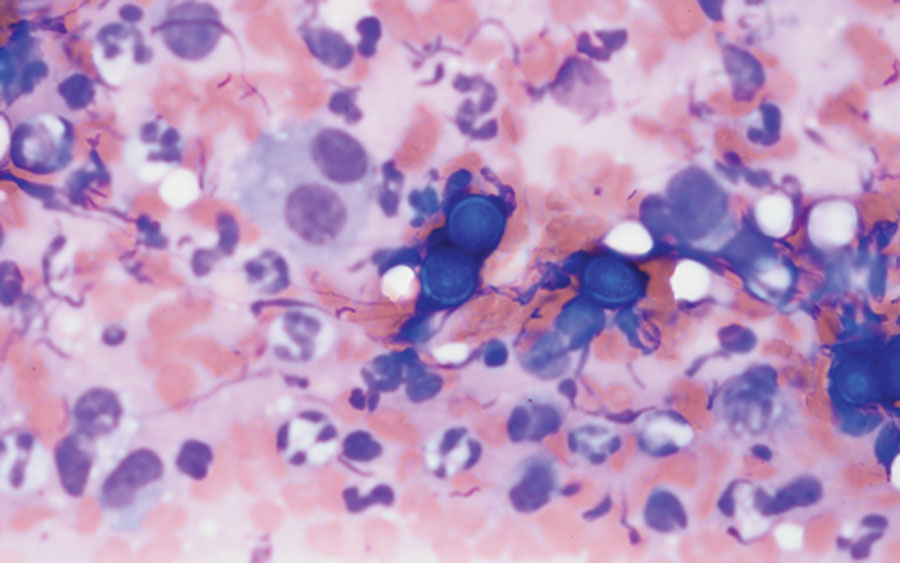
Canine Parvovirus Monoclonal Antibody
The canine parvovirus monoclonal antibody (CPMA) is a chimeric mAb that provides passive immunity when administered to dogs infected with canine parvovirus. It specifically targets the VP2 protein of canine parvovirus-2 (CPV-2), which facilitates entry into the host cell. Thus, CPMA neutralizes the virus by inhibiting cellular entry and prevents further tissues damage. Given this, CPMA may be more effective earlier in the course of the disease, but this has not been evaluated clinically. CMPA is a temperature-labile product and so is shipped and must be stored in specialized packing to prevent temperature fluctuations. Once defrosted, it is administered as a single 0.2 mL/kg IV bolus.
CPMA is conditionally approved by the USDA, which means that the product is safe and has a reasonable expectation of efficacy, but efficacy still needs to be evaluated in field studies. The initial data in purpose-bred animals are quite promising. In this study, 8-week-old puppies were experimentally infected CPV-2b and treated with CPMA or placebo 4 days post-exposure, at which point all dogs were showing clinical signs and positive on the fecal ELISA.
Without any additional treatment, 57% of the control dogs died compared with 0% mortality in the CPMA group. Additionally, dogs treated with CPMA had less severe clinical signs and laboratory abnormalities compared to placebo. Administration in healthy client-owned dogs is well tolerated with the most common adverse event being injection site reactions (4%).
Gilvetmab
Over the past decade, immune checkpoint inhibitors (ICIs) have revolutionized cancer therapy in humans. The healthy immune system has multiple mechanisms to regulate itself and prevent over-exuberant responses. One of these mechanisms is the expression of immune checkpoints, which are cell surface markers on lymphocytes that, when bound by their ligands, decrease lymphocyte activation. In the tumor microenvironment, cancer cells exploit this mechanism by increased binding of immune checkpoints to suppress the patient’s antitumor response and allow propagation of malignant tissues. ICI therapies, many of which are mAbs, block the immune checkpoint molecule or its ligand and thus reinvigorate the local immune response against tumor cells.
By Jennifer M. Reinhart, DVM, PhD, DACVIM (SAIM), DACVCP