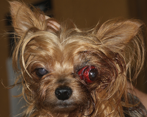
A proptosis refers to a sudden protrusion of the globe from the orbit. While they can appear quite scary, with some preparation they can easily be addressed. The number one question that you as a practitioner need to determine is: Should the globe be replaced, or should an enucleation occur? This injury may be caused by a multitude of traumatic situations, ranging from being attacked by a bigger dog to being hit by a car. Therefore, the pet’s overall needs have to be evaluated before the proptosis is addressed. During this stabilization period, you can treat the eye by administering systemic pain medication and applying topical lubrication. Once the pet is stable systemically, you can further evaluate the prognosis for the globe.
Indications for Replacement
Positive indicators include:
- Positive menace response (difficult to assess due to the eyelids being behind the globe)
- Positive consensual pupillary light reflex
- Fewer than 3 extraocular muscles ruptured
- Breed (brachycephalic breed has a better prognosis than a doliocephalic, such as a greyhound, collie, or dachshund)
Negative indicators include:
- Complete hyphema
- A corneoscleral laceration
- Optic nerve transection
- Corneal desiccation (drying)
- Species (dogs’ eyes can be replaced, cats’ eyes must be enucleated)
Proptosis Repair
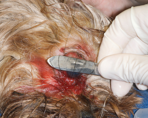
Once the decision has been made to replace the globe, prepare the patient for general anesthesia. After the patient has been anesthetized, use 3-0 or 4-0 silk to preplace two to three horizontal mattress sutures through stents, but do not tie. While doing this, ensure that the sutures are placed through the meibomian glands. Apply additional lubrication to the cornea and gently slide a bard-parker blade handle between the sutures and cornea.
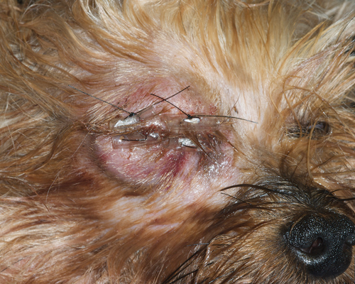
With an assistant holding the blade handle in place, lift up on all of the sutures at one time to facilitate the eyelids coming back over the globe. Tie all suture tails and send the patient home with a topical antibiotic and systemic pain medication. The tarsorrhaphy should remain in place for two to three weeks.
Potential Complications
to relieve tension.
The most common complications following proptosis are:
- Corneal ulceration
- Keratoconjunctivitis sicca (KCS)
- Strabismus
- Lagophthalmia
- Blindness
Take-home Points
The most important thing to remember is that while the proptosis may be the first thing that you notice in a trauma patient, it is not the medical priority. You need to ensure that the patient is stable systemically before addressing the proptosis. Additionally, lubrication early and often will aid you in preventing corneal ulcerations and allow you time to stabilize.
Finally, if unsure whether an enucleation or replacement should occur, inform the client of the risks. And remember: an enucleation can be performed at a later date, but you won’t get another chance to replace the globe.
By Todd Marlo, DVM, MS, DACVO
Reference
Miller, Paul E. “Ocular emergencies.” Slatter’s Fundamentals of Veterinary Ophthalmology, 4th ed. St. Louis: Saunders (2008): 419-426.