Although there have been many studies over the years on feline tooth resorption (TR), researchers have not determined the condition’s etiology. Proposed causes of TR include periodontal disease, feline immunodeficiency virus, feline leukemia virus, and feline herpesvirus-1. Studies, however, have proven inconclusive.
We do know that between 25% and 75% of the pet cat population in the U.S. is affected by TR. It can occur on any teeth, but it occurs most often on the buccal aspect of the premolar and molar teeth. TR tends to occur in cats over 2 years old and increases in prevalence as cats age.
Clinical Signs
Cats may have no clinical signs, especially during the early stages of the disease. When pulp exposure occurs, cats begin to exhibit ptyalism, head shaking, dropping food, and “chattering” of the teeth. As TR progresses, cats can become anorexic and lose weight.
During the anesthetized oral exam, the clinician should pay extra attention to the maxillary and mandibular third premolar teeth, the maxillary fourth premolar teeth, and the mandibular first molar teeth, which are commonly affected.
These teeth typically have granulation tissue covering the exposed pulp and bleed easily when probed with a dental explorer. If TR affects the canine teeth, they may appear to be super erupted and have peribulbar expansion on the labial side of the teeth.
Diagnostic Imaging Findings
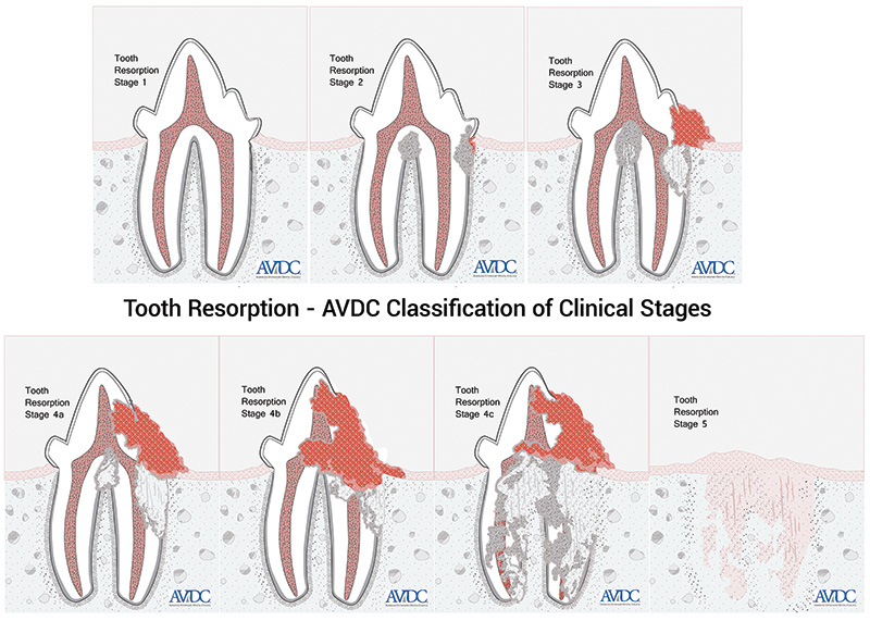
In addition to performing an anesthetized oral exam, the clinician must perform dental radiography to diagnose TR subgingivally and to decide on the best treatment plan for the patient. Radiographs allow determination of the stage and type of TR. TR stages 1 through 4 can be painful to the cat, so these teeth require treatment (Figure 1).
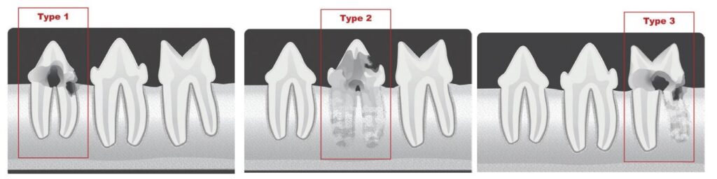
Radiographically, type 1 TR has a normal periodontal ligament space and radiolucencies in the tooth. Type 2 TR is characterized by replacement resorption, which occurs when the dental hard tissue is replaced by bone. Radiographs show a narrowed or nonexistent periodontal ligament space and radiolucency in part of the tooth. Type 3 TR is characterized by having type 1 and 2 resorption present in the same tooth (Figure 2).
Treatment for Tooth Resorption
Teeth with type 1 TR must be completely extracted.
A flap is first created to treat teeth with type 2 TR. A crown amputation is performed below the mucogingival line. FELV/FIV positive cats and cats with periodontal, endodontic, and periapical disease should not be treated with crown amputation.
Teeth with stage 5 TR do not require any treatment. Confirm that the overlying gingiva is intact.
A technique called “root pulverization,” performed on teeth with TR in the past, is not recommended due to potential serious adverse effects, such as trauma to soft tissues, neurovascular structures, and alveolar bone. Also, root fragments can be pushed into the mandibular canal, infraorbital canal, and nasal cavity.
References
Gorrel C. Tooth Resorption in Cats Pathophysiology and Treatment Options. J. Feline Med. Surg. 2015; 17: 37-43.
Reiter A, Soltero-Rivera MM. Domestic Feline Oral and Dental Diseases. In Veterinary Dentistry: Principles and Practice. Philadelphia: Lippincott-Raven Publishers, 2019.
By Suma M. Rao, DVM, MBA, Dipl. ABVP (Canine and Feline Practice)