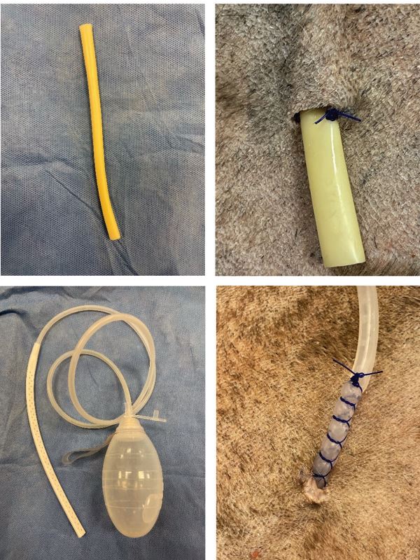
Examples of the Penrose drain (top) and Jackson Pratt drain (bottom) before and after placement.
By Clara Moran, DVM, MS, DACVS-SA
Drains are used commonly in small animal patients for a variety of indications: to address dead space, remove contaminated fluid, and improve tissue layer adherence. They are typically categorized into two groups: open passive drains and closed active drains.
Penrose Drain
A Penrose is an example of an open passive drain. These drains essentially act as a wick, keeping a small hole open adjacent to the incision to allow fluid to egress from the surgical site.
They are effective mainly due to the effect of gravity, capillary action as the fluid runs along the surface of the drain, and rising pressure within the wound bed.
To place the drain, create a small exit hole in the most gravity-dependent part of the wound bed, several centimeters away from the wound edge. It is important to position the drain appropriately, as this type of drain relies on gravity to encourage fluid to wick out.
Exit the drain from the wound bed through the hole, being sure that the skin is not too tight around the sides of the drain: if it is too tight, fluid will not be able to drain around it. Secure the drain by placing a single tacking suture through the drain and engaging the skin at the exit hole, leaving approximately 2 cm of drain exposed. The incision can then be closed routinely over the drain, being careful not to accidentally engage the drain while doing so.
It is typically not necessary to create separate entry and exit holes for Penrose drains. A dorsally positioned entry hole will typically not be responsible for much fluid egress, and may increase the risk of contamination of the wound bed. Once the wound is closed, a bandage should be placed to absorb discharge and provide mild compression to the surgical site.
Jackson Pratt Drain
A Jackson Pratt is an example of a closed active drain. This type of drain has a fenestrated end that is placed within the wound bed, then a section of tubing that is exited adjacent to the wound and connected to a reservoir.
Unlike Penrose drains, closed active drains can be exited wherever is most convenient, as they are not dependent on gravity to function. Instead, the reservoir itself generates negative pressure and pulls fluid out of the wound bed.
There are several different commercially available suction drains, but in the case of Jackson Pratts the negative pressure is generated by squeezing all the air out of the reservoir and closing the stopper. As the bulb slowly re-expands it will draw fluid in and collect it.
Just like for Penrose, the tubing for closed active drains should always be exited adjacent to the incision, never through it. Unlike Penrose, these drains should be secured with a pursestring and fingertrap, in order to maintain a tight seal of the exit hole around the drain and prevent air leakage, which will cause the drain to lose negative pressure.
In general, closed active drains should be emptied at least every 8 hours, or when the reservoir reaches half full (whichever comes first). They do not need to be bandaged unless it is necessary to secure it to the patient.
Pros and Cons
Closed active drains have several advantages over Penrose. As a rule, they tend to be more effective at removing wound fluid, thereby encouraging improved tissue plane adherence. The closed system limits wound contamination due to ascension of bacteria up the material of the drain. This also means it is safe to use a closed active drain in the abdomen, while Penrose should not be used for this purpose.
Additionally, accumulating fluid in the reservoir can be quantified and evaluated cytologically. Serial (usually daily) cytologies can allow the clinician to monitor the healing of the wound, as well as helping determine when the drain is ready to be removed.
While closed active drains are superior to Penrose in many aspects, Penrose can, when used appropriately, also provide a successful outcome and are considerably more economical than commercially available closed suction drains.

Make Your Own
An inexpensive way to create a closed suction drain that can be used in small wounds is to use a butterfly catheter. To create the drain, cut off the Luer lock on the end of the butterfly, then create small fenestrations in the distal quarter to third of the tubing. Be sure that none of these fenestrations exceed one-third the diameter of the tubing, or it could break off during removal.
The fenestrated portion of the tubing can then be placed within the wound bed just like a Jackson Pratt, and secured with a pursestring and fingertrap. This leaves the needle of the butterfly free; it can be inserted into a red top tube. The vacuum of the red top tube will draw fluid out of the wound, and the tube can be changed out as needed.
Caring for the Drain
Patients with drains should be monitored closely and must be restricted from accessing their drains. E collars, t-shirts, and bandages may all be used to prevent dislodgment or chewing the drain.
If a drain is broken or chewed off leaving part of it behind in the wound bed, the incision must be opened to retrieve it.
The drains should be cleaned regularly, and the bandage over a Penrose replaced every 1 to 2 days to prevent strike through.
As the wound heals, fluid production is expected to taper down and plateau, and serial cytologies should reveal a decreasing number of inflammatory cells and a shift from predominately degenerate neutrophils to more of a mixed population (mostly nondegenerate neutrophils, macrophages, lymphocytes).
Once these indicators are observed, it is appropriate to pull the drain. The drain hole should be left open to heal by second intention; it typically seals within 24 hours and until that time can be covered by a light adhesive bandage.
Wrapping Up
With correct usage, drains can aid in wound healing for many small animal patients. Drains are best used in minimally contaminated or sterile wound beds and should not be considered a replacement for open wound management in severely infected wounds.
Complications associated with drains are uncommon, but can include premature or incomplete removal, bacterial contamination of the wound, abscess formation at the exit site, and tumor seeding along the drain tract. Drains are not recommended in oncologic cases for this reason. However, in other contexts using a drain is one of the most effective ways to eliminate dead space, limit seroma formation, and allow complication-free healing.