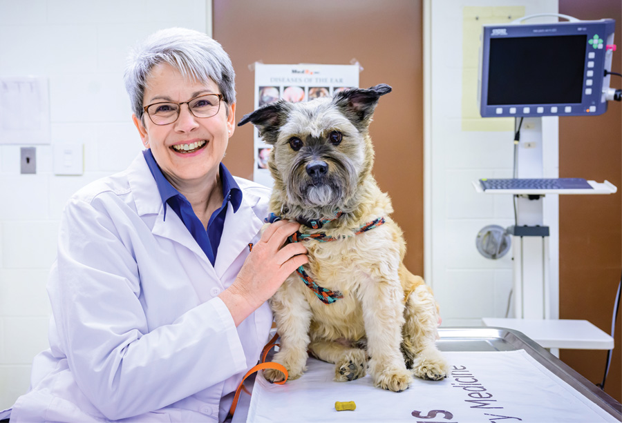
Dr. Clara Moran is a faculty member with the small animal surgery service.
Tell us about your background.
I’m from Georgia originally. I went to UGA for undergrad and for vet school, and have always maintained an interest in surgery. I matched to Illinois for internship, and found the college to be a great fit: full of lovely people and fostering a welcoming environment to develop my skills. I was lucky enough to stay on for a residency, which I completed in 2017. And since then, I’ve just never left!
How did you become interested in small animal surgery?
I started working for a veterinary surgeon in Savannah while I was in high school, and that made a huge impact on my career goals. He did a bilateral rostral maxillectomy with nasal planectomy the first summer I was there. He essentially took off a huge amount of a dog’s muzzle and then put everything back together! I was hooked; nothing else veterinary medicine had to offer ever seemed quite as cool.
What are your special interests?
I prefer soft tissue over orthopedics, and in particular enjoy challenging critical/emergent cases, such as major trauma cases. I like the intersection between surgery and critical care in those cases, and love working in synch with our ER and anesthesia departments to help pets that are severely ill or injured. I also like thoracic surgery and orofacial surgery; those tend to be some of my favorites. I also like wounds and reconstruction cases.
Tell us about one of your favorite cases.
Earlier this year a little dog presented to the hospital after being attacked by a stray while he was on a walk with his owners. Most of his wounds were centered around his face and neck. We did imaging and found that his hyoid apparatus was crushed, some of the bones of his skull were fractured, and he had multiple wounds penetrating into his oral cavity/pharynx.
We placed a tracheostomy and an E-tube and debrided his wounds. The worst of the wounds opened into a large pocket over his face, and the underlying tissues were badly lacerated or crushed.
It was a long procedure, but eventually we were able to remove everything devitalized and create a flap to help cover the area of skin loss. He stayed in the hospital for quite a while, but in the end everything healed beautifully. After the swelling in his pharyngeal region and larynx reduced, we pulled the tracheostomy and E-tube.
I took pictures along the way to document his progress, and the before and after is striking: from a gaping wound with ragged bits of muscle and loose bone fragments to a sweet old terrier face, with just a scar left to show what happened.


