Cutaneous masses—probably the most common structures aspirated cytologically—generally arise from one of three processes: inflammation, cysts, and neoplasia.
Fine-needle aspiration with cytology can help you determine what should be done next to attain a more definitive diagnosis or for diagnosis.
Inflammation
Inflammatory masses are very common and are categorized based on the type of inflammatory cells. Suppurative inflammation is predominated by neutrophils.
Pyogranulomatous consists of a mixture of neutrophils, epitheliod macrophages, and multinucleated giant cells. Lastly, granulomatous inflammation is heavily predominated by macrophages and multinucleated giant cells.
In a suppurative inflammatory response, if the cause is a bacterial infection, the neutrophils will often be degenerate and intracellular bacteria may be identified. Pyogranulomatous inflammatory responses often occur secondary to fungal infection or a foreign body response. Blastomyces dermatitidis commonly causes multiple masses or draining tracts and elicits a pyogranulomatous response.
Cysts
Cysts are epithelial-lined, non-neoplastic structures that are commonly seen in older dogs. Follicular cysts are often seen as dermal, raised, fluctuant masses that are filled with keratinaceous debris. Cytologically, thick aggregates of keratinaceous material and many anucleated, mature squamous epithelial cells are observed. These masses are benign, though, if they rupture, the free keratin can elicit a severe inflammatory response. The neutrophils and macrophages are often observed surrounding the keratinaceous material. Sebaceous masses, cysts, hyperplasia, and adenomas can look very similar.
Suggested Reading
- Raskin RE, Meyer DJ. Canine and Feline Cytology, A Color Atlas and Interpretation Guide, 2nd ed. Saunders Elsevier, St. Louis, MO. 2010.
- Cowell RL, Tyler RD, Meinkoth JH and DeNicola DB. Diagnostic Cytology and Hematology of the Dog and Cat, 3rd ed. Mosby Elsevier, St. Louis, MO 2008.
- Barger A. Common masses in dogs and cats. Clinician’s brief 7;9:9-11, 2010.
- Cohen M, Bohling MW, Wright JC, et al. Evaluation of sensitivity and specificity of cytologic examination:269 cases (1999-2000). J Am Vet Med Assoc 222:964-967, 2003.
Sebaceous cysts are less cellular than the other two and contain thick mats of nonstaining lipid-based secretory material. Low numbers of sebaceous epithelial cells may also be observed. These cells are cohesive. They have a moderate amount of basophilic cytoplasm filled with discrete cytoplasmic vacuoles. The nuclei are small and round but are often obscured by the vacuoles.
Sebaceous hyperplasia and sebaceous adenomas are cytologically indistinguishable. They are highly cellular and consist of multiple clusters of uniform sebaceous epithelial cells. Grossly, sebaceous adenomas appear as pedunculated wart-like masses, whereas sebaceous hyperplasia may appear as a thickened area of skin.
Neoplasia
There are many cutaneous and subcutaneous neoplasms that are diagnosed in dogs and cats, but this article focuses on the most common tumors.
Round cell tumors commonly appear as a red, raised hairless dermal mass. This grouping of tumors includes mast cell tumor, plasma cell tumor, lymphoma, histiocytoma, and transmissible venereal tumor. Of these tumors, mast cell tumors and histiocytomas are the most common.
Mast cell tumors are more common in dogs than cats. Cytologically, they are cellular and consist of a population of round cells with a moderate amount of basophilic cytoplasm, filled with metachromatic (purple) granules. These granules do not stain consistently with Diff-Quik and special staining may be necessary to clearly visualize these granules.
Eosinophils often accompany mast cell tumors in dogs but do so less commonly in cats. Well-differentiated mast cell tumors in cats usually do not contain eosinophils. Histopathologic assessment is necessary for grading in dogs, but grading has not proven useful in cats. In dogs the grading is important to attempt to predict the behavior of these tumors.
Histiocytomas are unique to dogs. They are benign tumors that will often resolve on their own after several weeks. Grossly they appear as a round, red, raised mass. Cytologically this tumor consists of a population of round cells with a moderate amount of basophilic cytoplasm and a centrally placed nucleus. The cytoplasm often has a clearing of the basophilia at the periphery. As the tumor begins to resolve, increasing numbers of small lymphocyte (cytotoxic T lymphocytes) can be observed, sometimes outnumbering the neoplastic cells.
Lipomas are a benign mesenchymal tumor and probably the most common benign tumor in dogs. Generally they are of low cellularity. Large droplets of lipid are observed on the unstained slide and often wash away during staining. Adipocytes are large cells with abundant, pale-staining cytoplasm packed with lipid and a small round nucleus. Benign spindle cells can also be observed in these samples. Lipomas, cytologically, are often indistinguishable from mature subcutaneous adipose tissue.
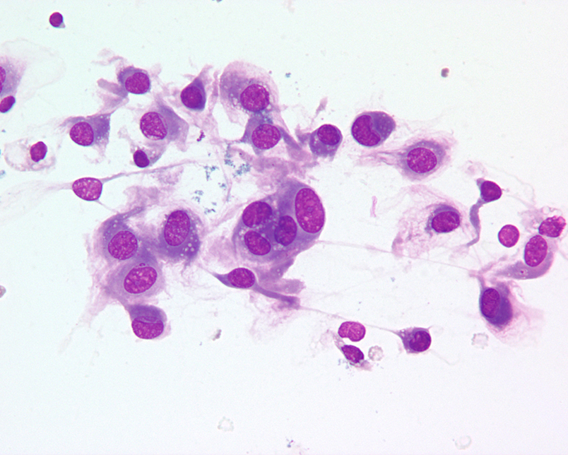
Squamous cell carcinomas (SCC) are a frequently diagnosed epithelial tumor. This tumor is the most common head and neck tumor diagnosed in cats. Samples often contain a mixture of cell types because the neoplastic squamous cells are frequently accompanied by inflammatory cells. The presence of inflammation can make diagnosis challenging because epithelium can become dysplastic secondary to inflammation. The neoplastic squamous epithelial cells are arranged in clusters with varying amounts of basophilic, occasionally keratinized cytoplasm. The nuclei are large and round with a stippled chromatin pattern and prominent nucleoli. Occasionally cells are coated in neutrophils. Grossly the masses are often ulcerated which contributes to the inflammatory component of the tumor. In conclusion, there are many benign and malignant processes that can be successfully diagnosed with cytology. The veterinarian’s clinical impression is critical in the appropriate interpretation of a cytologic specimen. Neoplastic processes are more likely to have a monomorphic population of cells, whereas inflammatory responses are more likely to be pleomorphic and painful to the patient.
By Anne Barger, DVM, MS, DACVP