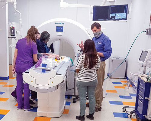
Above: Dr. Kenneth Welle directs the imaging of one of his patients in the 128-slice CT.
You know how much your clients care about their canine and feline companions, and that the demand for advanced diagnostics and therapies is on the rise. But you may not be aware that the same holds true for feathered, scaled, and miniaturized companions, too.
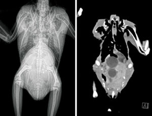
The zoological medicine service is seeing growth in referrals for advanced medical techniques. One of these is computed tomography, better known as CT. CT allows three-dimensional visualization of anatomy and allows us to more critically assess the clinical situation.
The scans are quite rapid and usually require only sedation. An intravenous or intraosseous catheter may be needed for contrast placement, but generally this imaging modality is entirely non-invasive. Often, the positioning is less stressful for the patient than traditional radiography. Although x-ray beams are utilized in this modality, the computer allows for the image to be rotated, the contrast to be adjusted, and other such manipulations to enhance visualization of various organs, lesions, or structures.
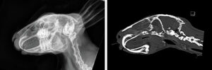
CT is particularly helpful in complex anatomical sites, such as the skull, where the many layers of overlapping bone can make interpretation very difficult. Likewise, large coelomic swellings in birds and reptiles can look like just one, large, soft tissue mass on radiographs, but we can distinguish individual organs on CT. The spinal column can be evaluated much more easily on CT than on radiographs. It can be very helpful in staging cancer, surgical planning, and any number of other scenarios.
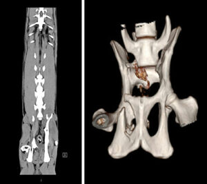
Rarely a week goes by without a few cases where we have made critical judgments based on CT. Some recent cases where we utilized CT include a budgerigar with a prolapse and distended coelom, where we discovered both a collapsed, shelled egg and an unshelled egg; a guinea pig with a rectal mass; another with a sialocele; and a parrot with chronic pulmonary interstitial fibrosis.
In some patients, CT is used to monitor progress. Some common indications for CT include dental conditions in rabbits and rodents, masses nearly anywhere on the body, and, in most species, surgical planning for uncommon conditions, spinal injuries, and orthopedic concerns. Geriatric birds can be evaluated for suspected atherosclerosis or pulmonary fibrosis, among other conditions.
It’s helpful when considering referral for CT that the referring veterinarian contact our service to discuss the case. We may be able to help decide if it would be the most beneficial modality for the patient and can discuss the process, the cost, and assist with scheduling.
Contact the Zoo Med service at vthzoomed@vetmed.illinois.edu.
By Kenneth Welle, DVM, ABVP (Avian)