![[Horse jumping]](https://vetmed.illinois.edu/wp-content/uploads/2021/04/warmblood-web.jpg)
Foot pain is a common presenting complaint in Warmblood horses worldwide. Our research group recently completed a large retrospective, cross-sectional study to determine the most common foot lesions diagnosed by magnetic resonance imaging (MRI) in Warmblood horses used for dressage, jumping, and eventing.
The study represented the largest population of Warmblood horses with foot pain ever studied. The subjects were 550 Warmblood horses that had been scanned using standing MRI of the distal limb(s) at B.W. Furlong & Associates Equine Hospital (New Jersey) or Advanced Equine Imaging of Wellington (Florida).
Data Collected
The following data were pulled from the medical records of these patients with foot pain: signalment, occupation, lameness, diagnostic analgesia, imaging results, treatments, and follow-up assessments.
Associations between foot lesions and chronic lameness following treatment were tested. All MRI images were interpreted by ACVR-certified veterinary radiologist Natasha Werpy, who has vast experience in equine MRI. Follow-up information was obtained from the medical records, veterinary recheck evaluations, follow-up MRI evaluations, and telephone interviews with the owners, trainers, or referring veterinarians.
Types of Lameness
One hundred and thirty-three horses (24.1%) had bilateral forelimb lameness, 379 (68.9%) had unilateral forelimb lameness, 15 (2.7%) had unilateral hind limb lameness, and 23 (4.3%) had no information recorded. The median lameness severity was grade 2 (on a scale of 1 to 4) while horses were trotted in a straight line and in circles on a smooth hard surface.
The lameness resolved on the lamest limb with analgesia of the palmar/plantar digital nerves in 367 horses (66.7%), or palmar/plantar nerves in 126 horses (22.9%), or distal interphalangeal joint (DIJ) in 57 horses (10.4%).
Imaging Abnormalities
Abnormalities of the navicular bone (409 horses, 74%), DIJ (362 horses, 65%), and deep digital flexor (DDF) tendon (260 horses, 47%) occurred with the highest frequency. Horses with recent lameness were more likely to become sound than were chronically lame horses.
The following abnormalities were statistically associated with chronic lameness following conservative therapy:
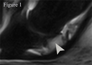
- Moderate to severe MRI lesions in the trabecular bone of the navicular bone (Figure 1),
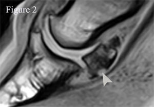
- Mild or severe erosions of the flexor surface of the navicular bone (Figure 2),
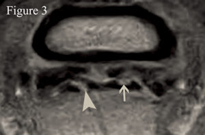
- Moderate sagittal/parasagittal DDF tendinopathies (Figure 3), and
- Moderate collateral sesamoidean desmopathies.
Also, identification of concurrent lesions of the DDF tendon, navicular bone, navicular bursa, and distal sesamoidean impar ligament was associated with chronic lameness after conservative therapy.
Athletic Demands May Contribute to Lameness
The most prevalent foot lesions detected in Warmblood horses used for jumping, dressage, or eventing involved the DIJ and/or the podotrochlear apparatus. Our findings suggest that the chronicity and severity of injury and the intended use of the horse play a major role in the response to conservative therapy.
We think that poor outcomes associated with certain foot injuries in this sample of Warmblood horses used for jumping or dressage activities could be due to the different athletic demands as compared to those previously reported for western performance horses.
Future research should be aimed to the development of effective treatment options for foot injuries in Warmblood horses that respond poorly to conservative or traditional therapies.
By Santiago D. Gutierrez-Nibeyro, DVM, MS, DACVS-LA, DACVSMR-Equine