It is more challenging than one may initially think to obtain radiographs that provide a diagnosis when patients come in for lameness. On top of needing more information, clinicians also have to consider the radiation exposure that their staff will receive while performing these films. Therefore, it is crucial that images be taken well, with the minimum exposure for both the patient and technical staff. Read on for tips and tricks that could not only improve the diagnostic quality of radiographs in your practice, but also provide a safer working environment for everyone.
Localization
The first and perhaps most important consideration before performing radiographs is lesion localization. Forelimb lameness is one of the more difficult things we see in orthopedics, and identifying the affected joint can be challenging. This is particularly true for the diseases affecting the shoulder and elbow.
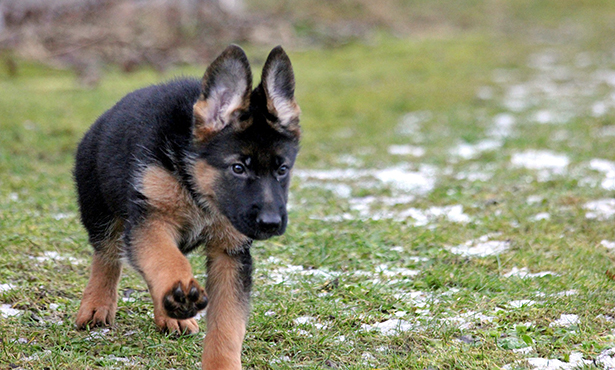
Once the source of pain has been identified, clinicians should request specific views that could highlight their suspected diagnosis. For example, if an ununited anconeal process in a 6-month-old German shepherd dog is at the top of the list, the flexed lateral elbow radiograph will be of the most diagnostic value. On the other hand, if a patient has biceps tendon pain on examination, it might be of more utility to perform an ultrasound of the tendon (or refer this patient to a facility with those capabilities).
Be advised that if cruciate pathology is suspected based upon physical examination and the patient is a candidate for TPLO (tibial plateau leveling osteotomy), the U of I orthopedics service will repeat radiographs for measurement and planning purposes, so it may be preferable in these cases to not perform radiographs (unless one is concerned for a bony lesion).
Sedation
The value of sedation for radiographs (or sometimes even a thorough orthopedic examination, depending on patient cooperation) is perhaps one of the more underutilized tools that we have. Sedation not only means that patients are less stressed during the procedure, but our technical staff can now step out of the room to take the film, minimizing their radiation exposure.
Most importantly, sedation allows us the time to position the patient exactly as we want them in order to obtain the most useful and diagnostic radiographs possible, which is what our owners are paying for. While a patient is under sedation, it is also an excellent time to confirm any findings you suspected on initial exam (cranial tibial thrust or cranial drawer) and assess for any laxity, if appropriate for that case.
For most healthy patients presenting for lameness, 0.2 to 0.3 mg/kg of butorphanol along with 4 to 6 μg/kg dexmedetomidine is appropriate for radiographic sedation. Reversal with an equal volume of atipamezole after study completion will typically allow the patient to leave the hospital walking within 15 to 20 minutes.
Orthogonal Views
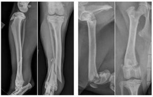
The importance of orthogonal views (lateral and anteroposterior) cannot be emphasized enough with regard to diagnosis. There are many occasions in which a fracture is visible in the sagittal plane but not the frontal plane, or vice versa. Similarly, it is equally important when taking post-operative films to perform orthogonal views to completely assess the repair or to assess healing during a subsequent recheck radiograph.
Interpretation
For challenging cases, there are teleradiology services that will provide a radiographic interpretation. It is always best to provide these radiologists with as thorough a history and physical exam findings as possible in order to have the most accurate reading.
Oftentimes a radiologist will see other lesions that are incidental, but the interpretation can be geared toward the patient if they have a precise history. The orthopedics service at the Veterinary Teaching Hospital is also always happy to consult on cases or help decide if a case should be referred for further work-up.
By Danielle Marturello, DVM, MS