![[lameness exam - gutierrez]](https://vetmed.illinois.edu/wp-content/uploads/2021/04/pc-lameness-gutierrez.jpg)
Veterinarians and Farriers Team Up on Lameness
Lameness. Technically, it’s defined as an abnormal gait of an animal. Practically speaking, lameness can mean the end of a sport horse’s career or a diminished quality of life for a pasture pony.
Because lameness is challenging to manage, it often requires a teamwork approach that includes a veterinarian and a skilled farrier.
Dr. Santiago Gutierrez-Nibeyro (in the photo above examining an equine patient) is an equine surgeon at the University of Illinois Veterinary Teaching Hospital. He says, “Veterinarians and farriers bring different perspectives to the problem of lameness.” Veterinarians use diagnostic imaging to see what is inside the foot, while farriers focus on what is outside.
The unusual case of bilateral keratomas in a six-year-old Holsteiner mare recently treated at the Veterinary Teaching Hospital illustrates how veterinarians and farriers work together from different angles to develop comprehensive treatment plans.
Curious Case of Keratomas
“Keratomas are a type of benign tumor that appear as a roughly circular mass, ivory to white in color,” says Dr. Gutierrez. “They are made of tough keratinized tissue and grow inside the hoof capsule, protruding into the underlying distal phalanx, the final bone in the hoof.”
This is uncomfortable for the horse and can cause apparent lameness.
The Holsteiner mare was referred to the hospital after 10 months of being intermittently lame in both front legs. Radiographs (x-rays) were inconclusive because the keratomas were located symmetrically on both feet.
![[lameness - custom shoe]](https://vetmed.illinois.edu/wp-content/uploads/2021/04/pc-hoofdetail.jpg)
Custom Shoe Plays Role in Surgical Procedure
It was determined that the best course of action would to be surgically remove the keratomas. A farrier and Dr. Gutierrez worked together to determine the best surgical approach and to develop a custom-made shoe to protect the hoof after surgery.
“A partial hoof wall resection was performed in each hoof centered over the toe using a Dremel rotary tool. The keratomas were easy to see after the hoof wall was removed,” explains Dr. Gutierrez.
“Then the tumors and adjacent tissue margins were carefully cut away. In addition, surface of the distal phalanx bone was debrided to ensure that all affected tissues were removed.”
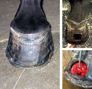
The surgical site was then thoroughly rinsed with antiseptic solution and packed with sterile gauze soaked with the same solution. The packing material was kept in place using stretchable elastic adhesive tape.
After the surgery, the mare was able to bear full weight on both front feet. Dr. Gutierrez reports that she showed some mild lameness in both front feet when walking, but this went away within a day.
Antimicrobial and anti-inflammatory medication was prescribed for the mare. The owner was instructed to keep both front feet wrapped and to change the wraps and packing every two days until the surgery site had dried and hardened. The mare also wore the special protective shoes.
Teamwork Ensures Optimal Patient Care
Thanks to the team effort involved, the outcome for this patient was successful. Both surgery sites healed without major complications. The mare regained full athletic function and returned to dressage competition, without the need for medication, within months of the surgery.
“Having the farrier and the veterinarian in the same room with the patient to discuss treatment plans and diagnostics eliminates confusion and ensures optimal patient care,” says Dr. Gutierrez.
If you have questions about therapeutic farriers and their contribution to lameness care, contact your local veterinarian or the Veterinary Teaching Hospital’s equine service.
By Hannah Beers