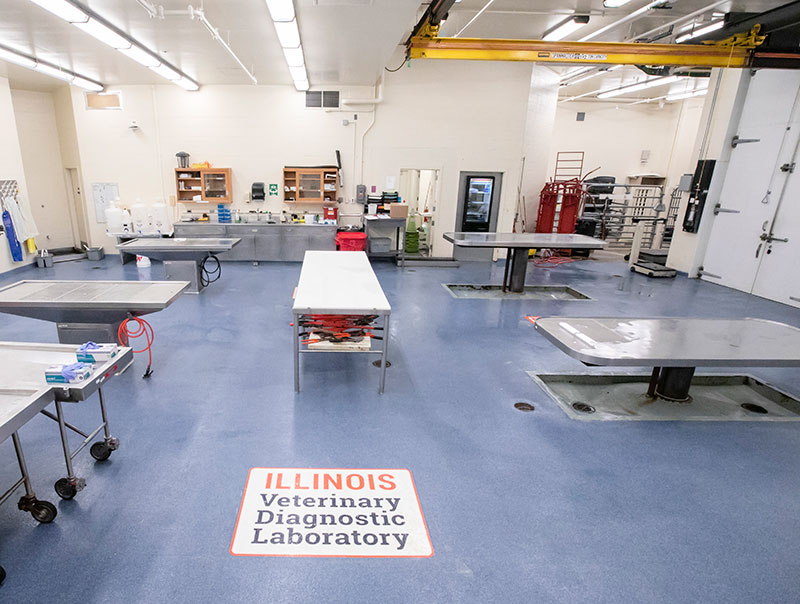
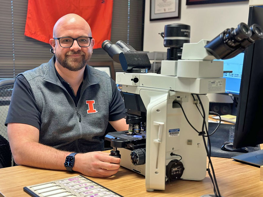
Like an autopsy in human medicine, a necropsy allows veterinarians to glean a wide range of information about a deceased patient. What steps are involved in performing a necropsy? And why would a necropsy be needed? Dr. Jonathan Samuelson, an anatomic pathologist at the University of Illinois Veterinary Diagnostic Laboratory, has the answers.
According to Dr. Samuelson, the necropsy consists of three parts: a gross examination, a histopathological examination, and supportive testing.
Using the Naked Eye and Microscope
The gross examination is when the animal and all its organs are observed with the naked eye. (This part of the necropsy is called gross because it is comprehensive, as in the phrase “gross income.” It is not “gross” in the sense of “blatant,” as in “gross negligence,” or in the sense of “disgusting.”)
During this portion, the pathologist is looking for any abnormalities, such as a change in color or texture. Some organs may be weighed and measured. Once an organ has been inspected and any abnormalities noted, tissue samples are collected for microscopic examination (known as histopathology) and any additional testing that may be required.
During the histopathology examination, the tissue samples collected during the gross examination are cut into thin sections. Then they are subjected to preserving chemical treatments and further sectioned into microns-thick sections. The final steps are to place them on a microscope slide and stain them. Staining allows the pathologist to see the cellular arrangement and structure of the tissue more distinctly. Abnormalities discovered during this process could indicate that an organ was not functioning properly, even if the organ looked normal during the gross examination. All the findings are recorded and added to those from the gross examination.
“Any given pathologist may put more weight on different aspects of the necropsy,” says Dr. Samuelson. “Some are more skilled at identifying and characterizing gross lesions and place more emphasis on these findings. Others use the gross examination of the body as a guide for more calculated microscopic examination and further testing.”
Further Testing
The final step of a necropsy involves additional testing. Experts at the Veterinary Diagnostic Laboratory have the capability to perform a huge variety of tests. For example, the microbiology lab performs aerobic and anaerobic cultures to find bacteria that may have caused disease.
The lab also uses MALDI-TOF mass spectrometry. (MALDI-TOF stands for “matrix-assisted laser desorption/ionization-time of flight.”) This molecular-based technology provides an even faster and more specific method for identifying microorganisms—mostly bacteria and some fungi—that cause disease.
“Another unit within the diagnostic laboratory, the virology lab, has the ability to perform virus isolation, antibody titers, and partial or whole viral genome sequencing,” adds Dr. Samuelson. These tests allow pathologists to confirm if the animal was exposed to or had a virus related to its death.
“Molecular testing may be performed with the aim of identifying a pathogen that might have contributed to the animal’s death,” Dr. Samuelson explains. The molecular lab performs PCR (polymerase chain reaction) analyses and other molecular-based tests to detect bacteria or viruses that could not otherwise be cultured or isolated.
Why Perform Necropsies?
Necropsies are performed for a variety of reasons. In some cases, when a pet’s death is unexpected, the veterinarian may ask the owners if they would like a necropsy done to find the cause of death. Law enforcement may ask for a forensic necropsy in cases of suspected animal abuse. Veterinarians serving food animal producers may recommend a necropsy to find out whether an unexplained death of an animal on the farm could indicate a problem within the herd that should be addressed.
Information gained in a necropsy may also help in the protection and management of wildlife. In 2022, a mountain lion in Illinois was hit by a car and killed. Because this species is not typically found in Illinois, personnel from the state’s natural resources department sent the body to the Veterinary Diagnostic Laboratory to be examined.
“In the case of wild animals, isotope testing can determine how old the animal is and where it came from,” says Dr. Samuelson. This information helps wildlife biologists understand the health status and migration habits of species across the country.
A necropsy provides a wealth of information. It may provide closure for pet owners or herd health information for animal producers. Necropsy findings may also isolate highly infectious disease organisms that pose a danger to animal and human health. These cases require immediate response by state or national agencies.
“Pathologists at the Veterinary Diagnostic Laboratory provide a valuable service,” concludes Dr. Samuelson. “Diagnosis is the business, but a pathologist does many other things as well. The information we gather can provide information, insights, and answers for future veterinary research and improving veterinary medicine.”
By Alaina Lamp