If only our dogs could maintain youthful agility their entire lives! Intervertebral disc disease is a degenerative process that is often associated with aging, though is most likely a multifactorial process. Dr. Lindsey Graham, a veterinarian pursuing specialization in neurology at the University of Illinois Veterinary Teaching Hospital in Urbana, offers information on identifying and treating disc disease. She also has suggestions for minimizing the risk of disc ruptures.
What Are Intervertebral Discs and Disc Disease?
An intervertebral disc is a structure located between most of the vertebral bodies along the spine. Dr. Graham compares the normal intervertebral disc to a jelly doughnut: it has a soft inside (called the nucleus pulposus) and a tough outer layer (called the annulus fibrosus).
“Intervertebral discs absorb shock, evenly disperse pressure along the spinal cord, and allow the spinal column to move fluidly when our pets are performing their daily activities,” she explains.
As our pets age, the intervertebral discs begin to degenerate. Intervertebral disc disease (IVDD) can be categorized into two types.
“Type I is most frequently associated with smaller breed dogs with short legs and long backs,” says Dr. Graham. Breeds that fall under this description include dachshunds, beagles, French bulldogs, and Pembroke Welsh corgis. However, Dr. Graham notes, any breed can be affected by this condition.
In Type I IVDD, the nucleus pulposus dehydrates and hardens, and the annulus fibrosus becomes thinner. When enough motion and force are exerted on the degenerating disc, the nucleus pulposus can herniate (or extrude) through the annulus fibrosus, leading to a defect in the annulus fibrosus and varying degrees of spinal cord compression.
In Type II IVDD, the nucleus pulposus degenerates and the inner layers of the annulus fibrosus lose their integrity, but the outer layers of the annulus fibrosus remain intact. This leads to chronic (slow) protrusion, a process in which the entirety of the intervertebral disc gradually pushes upward into the spinal cord. This condition is most commonly associated with larger breed dogs, but similar to Type I, any dog breed and size can be affected.
How Is Disc Disease Diagnosed?
The gold-standard for diagnosing IVDD is magnetic resonance imaging (MRI).
“MRI gives us the ability to determine the exact location of the degenerated or herniated (extruded) intervertebral discs,” says Dr. Graham. “MRI also gives us the ability to visualize the spinal cord and determine the degree of spinal cord compression and tissue damage (such as bleeding) caused by the intervertebral disc herniation.”
Another imaging modality, computed tomography (CT), can also be beneficial in diagnosing Type I IVDD, especially in patients whose intervertebral discs tend to mineralize as part of the degenerative process.
“Since CT is a great tool for evaluating bone and mineralized disc material is similar in appearance to bone, we are also able to visualize this material,” says Dr. Graham.
CT also provides information regarding the location of the affected intervertebral discs. MRI, however, gives veterinarians a clearer picture of the extent of the damage to the spinal cord than does CT. Also, if the patient’s intervertebral disc material has not mineralized, a CT scan might not detect the problem area.
Myelography, which involves injecting a dye within the layers of the spinal cord before obtaining radiographs or CT, is another imaging tool used to diagnose IVDD. Myelography is less commonly used compared to a standard CT or MRI, which often provide superior detail with fewer complications.
Radiographs (x-rays) of the spine may reveal obvious signs of intervertebral disc disease. To determine the extent and significance of abnormalities noted on radiographs and to plan for potential surgical intervention, however, requires advanced imaging (myelography or, more commonly, CT or MRI).
What Are the Treatment Options?
There are two main treatment options for intervertebral disc disease.
Conservative management consists of 4 to 6 weeks of strict crate rest, meaning that the patient is confined to a crate, playpen, or small room except when walked on a leash outside to urinate and defecate. Dr. Graham stresses that patients should not be allowed to run, jump, use stairs, or play with other pets during this period. To help facilitate rest, veterinarians might prescribe an anti-anxiety medication.
“Strict rest is truly the most important aspect of conservative management as it allows any defects in the annulus fibrosus to form scar tissue, which often prevent further herniation of intervertebral disc material and further spinal cord compression,” says Dr. Graham.
In cases where conservative management is unsuccessful or the patient is severely affected, decompressive surgery is recommended. The goal of surgery is to alleviate the compression of the spinal cord by removing the herniated intervertebral disc material.
“We typically reserve surgical intervention for patients who are unable to walk, are experiencing pain that cannot be adequately controlled with medications, or have not improved with conservative management,” explains Dr. Graham.
Recommendations
Unfortunately measures to prevent degeneration of intervertebral discs have not been identified. There are, however, steps pet owners can take to minimize the risk of intervertebral disc herniation and subsequent spinal cord compression.
“We recommend limiting jumping activities,” says Dr. Graham. “Running up and down stairs, jumping on and off furniture or other heights, uncontrolled off-leash activities, and roughhousing with other pets have been associated with an increased risk of intervertebral disc herniation.”
Installing doggy ramps can reduce risky behaviors for pets accustomed to jumping on furniture. Dr. Graham also recommends that pet owners use a chest harness rather than a neck collar to walk high-risk breeds or dogs previously diagnosed with IVDD. A harness more evenly disperses the pressure along the spine and protects the intervertebral discs in the neck.
“We encourage slow, controlled walks for pets at risk,” says Dr. Graham, “and we strongly advise minimizing the pet’s ability to engage in running and jumping.”
By Crystal Munguia
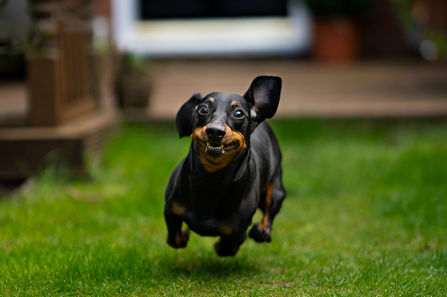
Photo by James Watson on Unsplash