Normal aging is usually associated with a decline in memory, although it is unclear what factors play a role. In a new study, researchers studied specific interneurons, which serve as communication centers that connect other neurons, in the regions of the brain that are important for learning and memory.
Increasing age places people at risk, whether it is because of a normal decrease in cognitive ability or due to postoperative cognitive disorders. In the latter, the deficits can persist for many months after surgery especially when the patients are older than 60. Unfortunately, the underlying cellular mechanisms that cause these impairments are largely unknown.
Previous studies have shown that the region of the brain that is associated with learning and memory—the hippocampus—decreases in volume with age. Additionally, the levels of the molecule y-aminobutyric acid, or GABA, and some of the interneurons that release it are also affected.
In the present study, the researchers focused on the hippocampal interneurons in a specific region, called the hilus of the dentate gyrus, that are characterized by their expression of somatostatin. This hormone has the ability to counteract the effects of growth hormones elsewhere in the body.
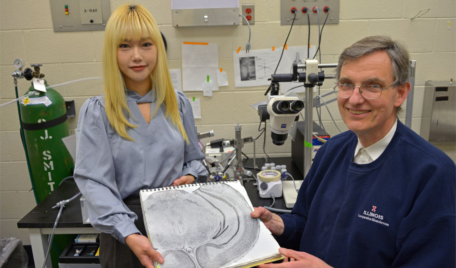
“In the past, other researchers have found that one of the differences between cognitively impaired and unimpaired rats was that the former had a lower number of somatostatin-positive interneurons in the hilus of the dentate gyrus,” said Uwe Rudolph (GNDP), a professor of comparative biosciences. “We wanted to further investigate whether a loss of these neurons is really responsible for cognitive deficits, and whether it could thus serve as a model of aging in the hippocampus.”
The researchers decreased the numbers of somatostatin-positive interneurons in the hippocampus of mice by using a toxin. They injected this toxin into the dentate hilus, so that the toxin would only be expressed in the somatostatin-positive interneurons, killing approximately 50% of these cells. Starting 3 weeks later, they conducted behavioral studies to test the learning abilities and memory of the mice.
The mice underwent three types of tests: whether they could remember and differentiate novel objects from familiar ones, navigate mazes using their short-term memories, and use their spatial learning to find a hidden platform in a pool of water. In all three cases, the mice that had decreased levels of somatostatin-positive interneurons struggled compared to those that did not receive the toxin.
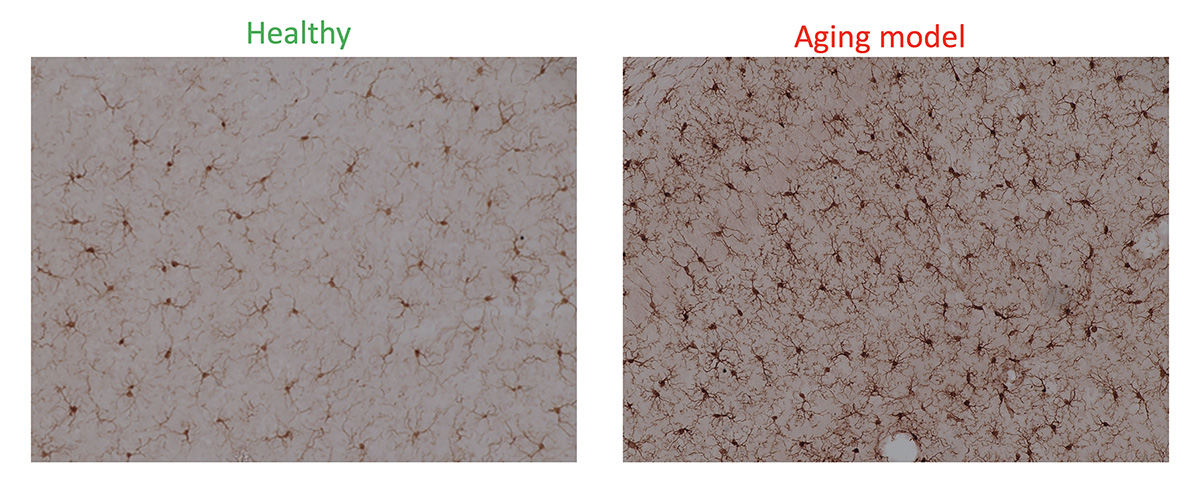
The researchers also looked for changes in cellular signals that occur due to lower levels of somatostatin-positive interneurons. To do so, they focused on the microglia, which are immune cells that are among the first to respond when something goes wrong in the brain.
“We looked at microglial activation, which is a hallmark of the inflammation that is associated with aging and memory impairment,” said Rajasekar Nagarajan, a postdoctoral researcher in the Rudolph lab. “We saw increased activation of microglia in the hippocampus of the mice that were injected with the toxin, even outside of the dentate gyrus.”
In addition to determining the status of microglia, the team measured a protein called brain-derived neurotrophic factor, which is active in the hippocampus and plays a role in long-term memory. They found that there were lower BDNF levels in the hippocampal tissue of the toxin-injected mice. Furthermore, the researchers also found that these mice had fewer hippocampal dendritic spines, which are critical for learning and memory.
Unsurprisingly for the researchers, the results seen with the toxin-injected mice were essentially the same as with aged mice that were 18-19 months old and had not been injected with the toxin.
“18 months-old mice correspond to an age of approximately 60 years in humans. It roughly fits the time point at which we know that people more frequently develop neurocognitive deficits in response to surgery and anesthesia,” Rudolph said. “Our results have shown that using this toxin to reduce the number of somatostatin-positive interneurons is sufficient to cause effects that resemble cognitive deficits in aging.”
The researchers are excited that they can use these techniques to investigate aging-related defects without waiting for the mice to grow old, which is an expensive and time-consuming undertaking. “We will be using this method as a model to test which experimental compounds can be used to prevent or reverse both age-related and post-operative cognitive impairments,” Nagarajan said.
By Ananya Sen, Carl R. Woese Institute for Genomic Biology
Originally posted here: https://www.igb.illinois.edu/article/specific-interneurons-are-important-aging-associated-cognitive-decline-study-finds
The paper “Genetic Ablation of Dentate Hilar Somatostatin-Positive GABAergic Interneurons is Sufficient to Induce Cognitive Impairment” was published in Molecular Neurobiology and can be found at https://doi.org/10.1007/s12035-023-03586-3. The study was funded by the NIH.