When an immunocompromised person’s system begins to recover and produce more white blood cells, it’s usually a good thing – unless they develop a potentially deadly inflammatory condition. New research from the University of Illinois Urbana-Champaign has found that the pulmonary distress often associated with the condition is caused not by damage to the lungs, but by newly populated T-cells infiltrating the brain.
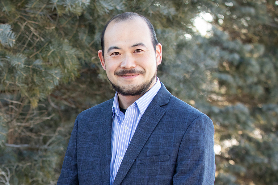
Knowing this mechanism of action can help researchers and physicians better understand the illness and provide new treatment targets, said study leader Makoto Inoue, a professor of comparative biosciences at Illinois. The study was published in the journal Nature Communications.
C-IRIS Affects Chemo Patients, Other Immunocompromised
Cryptococcus-associated immune reconstitution inflammatory syndrome, known as C-IRIS, happens when an immunocompromised patient is unknowingly infected with the fungus Cryptococcus. Once the patient’s immune system begins to rebuild, creating more T-cells, the infection sparks systemic inflammation.
C-IRIS frequently affects patients receiving antiretroviral therapy, recovering from chemotherapy, or recovering from a transplant, and also has been known to affect postpartum women or patients with multiple sclerosis – yet it remains difficult to diagnose, requiring ruling out other causes first, said study co-author Jinyan Zhou, a graduate student researcher at Illinois.
Wanting to understand more about the condition and its progression, the researchers developed a mouse model of the disease. To achieve this, they give injections of T-cells to immune-deficient mice preinfected with Cryptococcus, simulating what happens when the immune system starts producing higher levels of T-cells after being suppressed. The symptoms the mice developed – such as inflammation, fluid in the brain and pulmonary dysfunction – were in line with those of human patients.
‘T-Cells Infiltrating the Brain’
“We saw a high number of T-cells infiltrating the brains in conjunction with the presence of pulmonary dysfunctions, so that told us they could be connected,” Zhou said. “In healthy conditions, there shouldn’t be that many T-cells in the brain, because those cells should primarily exist in the periphery with a small number of T-cells doing patrol and surveillance within the brain.”
When the researchers investigated further, they uncovered a chain of events leading the T-cells to invade the brain and affect breathing. The receptor CCR5 is implicated in HIV and cancer, and also found on the surface of T-cells. When the T-cell populations started to rise in the mice, the receptor promoted the white blood cells’ infiltration into the brain. In addition, the T-cells that had infiltrated the brain produced high amounts of two molecules known to cause damage to neurons in the regions of the brain that control respiratory function.
“Finding that the pulmonary dysfunction is due to neuron damage in the brain was a new way of seeing this condition,” Inoue said. “We know the brain controls may peripheral organs, but this is a notable idea for clinicians treating this syndrome. Normally, drugs are given to try to improve lung function, but they don’t work. Now we know it’s due to changes in the brain, so that gives us new treatment pathways to target.”
Potential Treatments
To verify this chain of events and identify potential treatment pathways, the researchers treated a set of mice with a drug that suppresses the CCR5 receptor. Both pulmonary function and neuronal development improved.
“We think this could have potential to be beneficial for C-IRIS patients as well, to prevent T-cell infiltration into the brain, and that in turn could prevent the symptoms from happening,” Zhou said. “We also could target those molecules that damage the neurons. For example, when patients are prescribed immunotherapies we could maybe manipulate those T-cells to decrease their expression of those molecules.”
Next, the researchers plan to further study other functions of the immune system in C-IRIS and how more systems in the brain and body interact with T-cells.
The National Institutes of Health supported this work via grant R01-AI136999.
By Liz Ahlberg Touchstone, Biomedical Sciences Editor, Campus News Bureau
Featured photo by L. Brian Stauffer