In a paper recently published in PNAS Nexus, researchers from the Department of Comparative Biosciences at the University of Illinois Urbana-Champaign describe how they were able to inhibit the activity of a specific neuronal cell type in the hippocampus to induce cellular and behavioral changes associated with aging. Their study confirms a causal relationship between reduced function of certain interneurons in the brain and cognitive decline. In addition, their novel experimental model may be useful for studying age-related cognitive disorders.
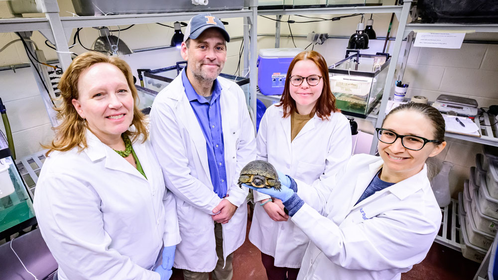
First author Jinrui Lyu, a graduate student in the Neuroscience Program and the Department of Comparative Biosciences, performed and analyzed the experiments for the study. Corresponding author Uwe Rudolph is professor and head of the Department of Comparative Biosciences in the College of Veterinary Medicine.
These authors answered questions about the design and significance of their study.
What was the motivation for your study?
Our lab is interested in why the aging brain is more susceptible than the younger brain for impairments of cognitive functions in response to surgery, which can last for several months and are associated with increased morbidity and mortality. We want to better understand what’s happening at a cellular level so we and others can ultimately develop therapies to treat this clinical problem.

In the present study, we wanted to test whether a reduction in functionality of a specific subset of neurons is responsible and sufficient for age-related cognitive decline, and to develop a new mouse model for aging in the hippocampus, a part of the brain that plays an important role for learning and memory.
It was known that the number of somatostatin interneurons in a subregion of the hippocampus, the dentate gyrus hilus, is reduced in aged cognitively impaired rodents. We tested the relevance of this observation by using chemogenetic tools to inhibit just these neurons. If this chemogenetic inhibition would lead to cognitive decline, this would establish a causal relationship between a decrease in the functioning of these neurons and age-related changes in cognition. This hypothesis was confirmed.
Because aging is a chronic process, we compared chronic inhibition of these somatostatin neurons (over at least 3 weeks) with more subtle intermittent inhibition, and in line with our expectations chronic inhibition had a much larger effect.
Aging is an inevitable, complex, and multifactorial process. … Understanding the cellular mechanisms underlying aging is crucial to develop novel strategies to prevent or to reverse age-related cognitive dysfunction.
What’s unique about this mouse model for aging?
In addition to cognitive impairments, we found several other changes that have been described in the aged brain. These include hyperactivity in the dentate gyrus, a reduction in the amount of somatostatin in the dentate gyrus (most likely due to the inhibited cells no longer expressing this marker), and increased microglial activation. Microglia are the resident immune cells of the brain and respond to changes in their environment.
The demonstration that the modulation of just this small cell population is sufficient to induce changes seen with normal or chronological aging in a part of the brain is what is novel. Aging is certainly associated with a multitude of changes in the brain—known and unknown—and it is striking that a minor change like the one we made is sufficient to explain several molecular and behavioral changes seen in aged mice.
One could also say that we found a way to make young mice (at least their hippocampus) old in a matter of just a few weeks. That was actually our goal, as we are also studying cognitive dysfunction of the aging brain in response to surgery. Working with aged mice poses significant logistical challenges, and we and others can now study the effects of aging in the hippocampus in chronologically young adult mice.
Can you explain what interneurons are? And what does “chemogenetic” mean?
Interneurons carry information between two other neurons, e.g., principal neurons. They inhibit and thus fine-tune and sculpt the activity of excitatory neurons. Interneurons likely play a role in all higher brain functions, including learning, memory, and planning. While estimates vary, both for different brain regions and also between species, it is widely assumed that in humans 20% to 30% of all neurons are interneurons, which can be classified into an almost countless number of subgroups, based largely on proteins they express, like somatostatin in the neurons that we were studying.
Chemogenetics involves the use of genetically engineered receptors that respond to specific small molecules that ideally do not have other effects. These receptors are packaged into viral vectors which are stereotaxically injected in the brain region of interest, where they will be expressed only in defined cells.
We chose a chemogenetic receptor that inhibits essential biochemical cellular pathways so that when we give a very low dose of the antipsychotic drug clozapine, only the cells expressing the chemogenetic receptor—in our case the somatostatin interneurons in the dentate gyrus hilus—are inhibited.
While we have not done such experiments, one could also test whether aging-related changes induced by inhibition of these neurons are reversible, by stopping the administration of clozapine.
How did you determine whether the changes you made in the mice affected their brain cells and their cognitive function?
We for example determined the density of dendritic spines, which are small protrusions from a neuron’s dendrite and which regulate the amount of excitatory neurotransmission and are thus of functional importance, e.g., for learning and memory. We also found that the cell body size of microglia was increased, consistent with microglia being activated.
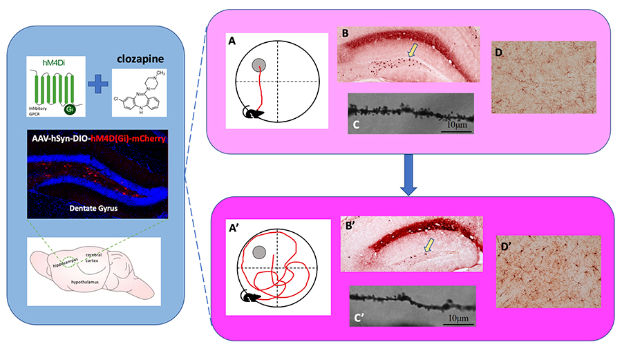
For behavioral analysis, we performed a variety of tests that provide information on different aspects of learning and memory. For example, we tested whether mice display the usual alternation behavior, i.e., enter into arms most recently not visited when they are allowed to freely move in a Y-shaped maze, or prefer to interact with a novel object over a familiar object. In the Morris water maze test, mice are trained to find a hidden platform, and when we move the platform to a new position, we are studying reversal learning, which has actually been linked to the dentate gyrus previously.
Why is this study important?
Aging is an inevitable, complex, and multifactorial process. Cognitive decline, such as memory loss, is one of the most prevalent concerns in the aging population. The hippocampus plays a vital role in learning and memory in the mammalian brain. Understanding the cellular mechanisms underlying aging is crucial to develop novel strategies to prevent or to reverse age-related cognitive dysfunction and to prevent or reverse postoperative cognitive deficits that occur primarily in aged patients.