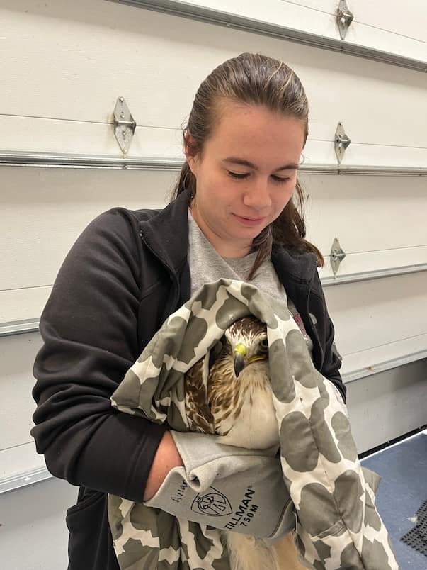
Follow Tyson Jenkins, WMC senior manager, and Alyssa Guest, WMC junior manager in training as they discuss their role in the Wildlife Medical Clinic, what their plans are after vet school, and how volunteering in the clinic will help them with those plans.
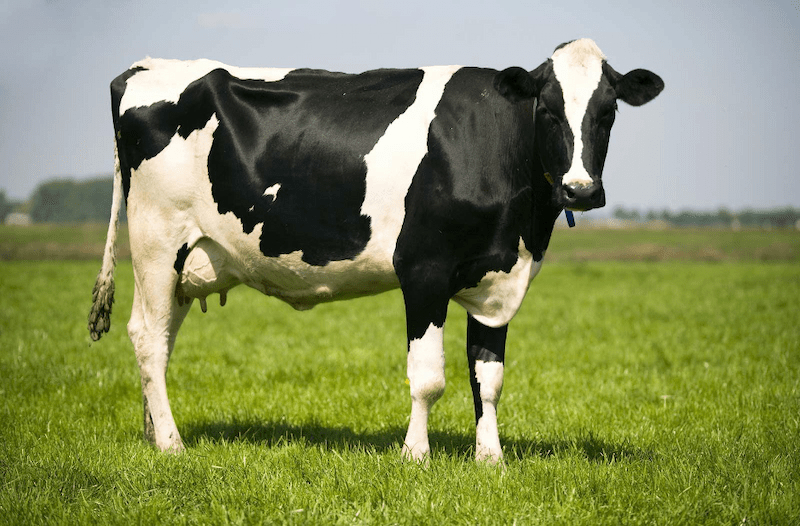
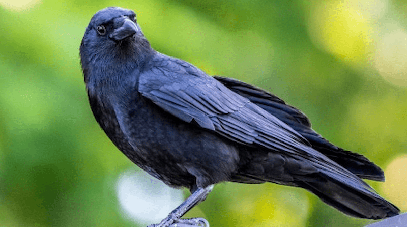
Highly pathogenic avian influenza, commonly referred to as HPAI, has become a common topic in conversation today. This may be attributed to the virus being detected in animals other than poultry or wild birds, including various mammalian predators and dairy cows, as well as several large avian mortality events attributable to the virus.
Avian influenza had previously been undetected in dairy cattle, but a discovery in 2024 identified the virus on certain farms. While the risk to public health is low at this point, preventing exposure is imperative in reducing opportunities for the virus to mutate.
Preventing Infection
The IDNR and IDPH urge the public to avoid physical contact with any waterfowl or any wild birds showing signs of illness. Pets can be infected by contact with carcasses, respiratory secretions, or droppings from infected animals, so it is important to keep your pets indoors or leashed and ensure they do not approach any sick or deceased animals.
Pets and humans can also be infected if consuming underdone infected poultry or raw milk from an infected cow. Being diligent to properly cook your food and avoid feeding your pet raw food or milk can prevent infection. If you have backyard poultry, keep them in a covered enclosure to prevent wild bird feces from dropping into the enclosure. Additionally, change your clothes and shoes before interacting with your poultry, and keep rodents and wildlife away from their food and water.
Deceased Wildlife
It is encouraged to avoid handling bird carcasses, but in the event carcasses must be removed from your property, there are steps you can take to protect yourself. If you come across a die-off of five or more birds you should contact your IDNR district wildlife biologist. When handling a deceased bird, you need to protect yourself by wearing the correct equipment. This includes waterproof gloves, safety goggles, an N95 mask, a hair covering, shoe covers, and a disposable gown or coveralls to protect your clothes.
Once you have your protective equipment on, you can dispose of the carcasses in a secure plastic bag and double bag the carcasses. Disinfect the outside of the bag with a disinfectant labelled for the flu virus and place it in a secure garbage can where animals will not get to the carcasses. When you are finished, carefully remove the protective equipment without contaminating yourself or clothes, double bag the equipment and again disinfect the outside of the bag and dispose of it in a waste container.
If you are unsure of how to remove your protective equipment, the CDC has information on how to safely do so. Make sure to wash your hands thoroughly afterward and avoid touching your face during the process.
Monitor for Exposure
If you have come into contact with a sick or deceased bird, even if disposing of a carcass with the correct equipment, you should contact your local health department, who will assess your exposure and help with symptom monitoring. Monitor yourself for flu-like symptoms after exposure and contact your health department if you develop symptoms up to 14 days after exposure. If you are experiencing symptoms and suspect you were exposed, do not show up to your healthcare provider without contacting them and the health department.
References:
Centers for Disease Control and Prevention. (n.d.). Interim guidance for employers to reduce exposure to avian influenza A viruses for people working with animals. Centers for Disease Control and Prevention. https://www.cdc.gov/bird-flu/prevention/worker-protection-ppe.html
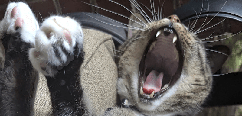
The Wildlife Medical Clinic has indeed seen it all in terms of injuries from human, domestic animal, and wildlife sources. Whether it be an accident or by instinct, all injuries, big and small, can become lethal to an animal if not adequately treated. One source in particular can be silent but deadly, wounds from a cat’s mouth.
A wound from a cat bite can harbor up to thirteen bacterial species from feline saliva. Most bites can host a mixture of multiple aerobic and anaerobic bacteria that can worsen puncture wounds and prevent healing or lead to sepsis. Several studies discovered that Pasteurella multocida is the most common pathogen within cat bites and is known to accelerate infections. At the Wildlife Medical Clinic, bite wounds are treated immediately with antibiotics and monitored very closely for any sign of infection that can indicate a difference between a healthy release and sepsis.
Case Correlation
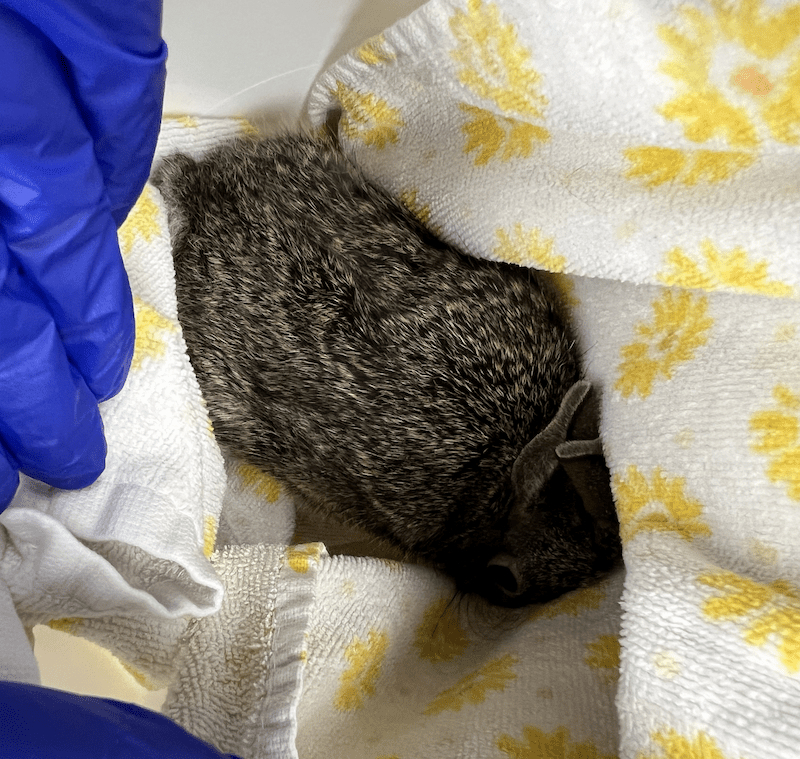
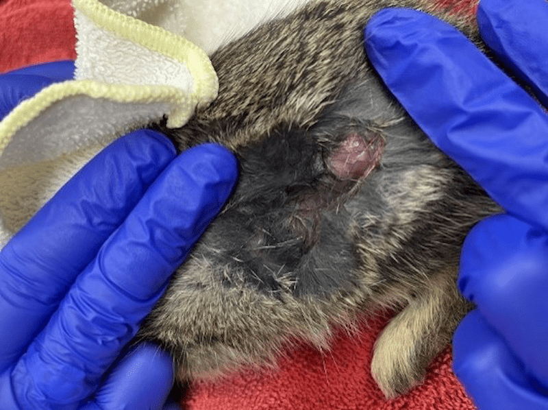
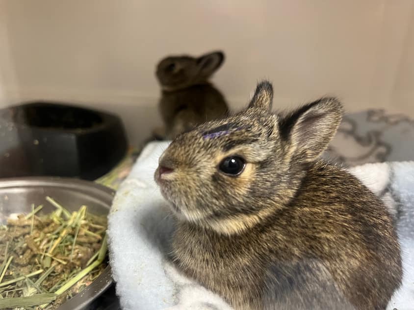
The Wildlife Medical Clinic recently treated a juvenile eastern cottontail after it was bitten by a cat. Upon examination, a large 1.5 inch by .5-inch de-gloving, which is a wound where skin is pulled off the underlying muscle and tissue. Also, a wound was found over the left stifle (knee), but it did not appear to enter the abdominal cavity.
Because of the very thin and friable nature of wild cottontail skin (imagine wet tissue paper), it was decided that any attempt to clip, clean, or suture the wound could cause further damage. Additionally, bite wounds should not be sutured closed, as that can trap dangerous bacteria under the skin. A simple flush of the wound was performed to remove excess debris.
A few other small abrasions were noticed but ultimately were of no concern. The eastern cottontail was ambulating (moving around) appropriately, was of a healthy weight, and had only mild dehydration. We decided that the best course of treatment was to provide fluids, pain medication, and antibiotics. To minimize stress and worsening of the wound, we were hands off as much as we could be with this patient.
The Outcome
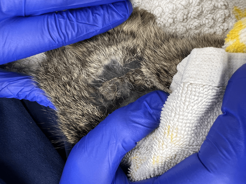
Young rabbits can heal their wounds with impressive speed, and several days later, the wound began to show signs of healing. With no signs of infection or pain, antibiotics and pain medications were discontinued from daily treatment with the plan to continue to monitor. Within eight days, the wound was fully scabbed and posed no threat to normal movement. The eastern cottontail was of healthy weight and was cleared for release! Another successful and rewarding case at the Wildlife Medical Clinic at the University of Illinois!
References:
Talan, D. A., Citron, D. M., Abrahamian, F. M., Moran, G. J., & Goldstein, E. J. C. (1999). Bacteriologic analysis of Infected Dog and Cat bites. New England Journal of Medicine, 340(2), 85–92. https://doi.org/10.1056/nejm199901143400202
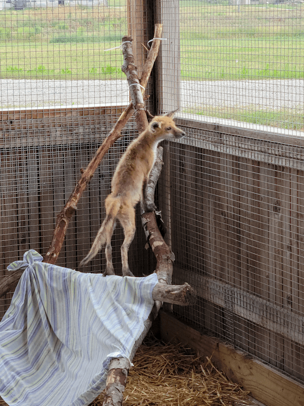
The red fox (Vulpes vulpes) is a part of the Canidae family and can be found to weigh seven to eighteen pounds, which is about the same weight of the average house cat! They also come in a range of sizes but on average these animals are about three feet long from their nose to the tip of their fluffy tail!
Red foxes are found all throughout Illinois but are less commonly seen in the southern part of the state. They are becoming more common in urban communities due to being outcompeted and predated by growing coyote populations. Foxes are mostly active at night which means they are nocturnal! However, they can also be crepuscular, where you may see them wandering around at dawn and dusk. This is prime time to go hunting for small mammals such as mice and rabbits.
While these animals may appear to be a carnivore, they are actually omnivores! Aside from eating small animals, come summer time, they also dabble in snacks such as local fruit/berry plants, vegetables, other edible plants, and insects.
The Dangers they Face
Due to habitat loss and the increase in coyotes, foxes have had to start relocating to more urban locations. While this is not ideal, they have managed to adapt. However, humans are one of their greatest threats. One of the largest causes of mortality is collisions with vehicles. Aside from that, hunting and trapping of fox is another danger they face.
The average lifespan of a wild red fox is about three to seven years. At birth, birds of prey (falcons, hawks, owls) are another source of mortality, specifically in kits (young foxes). Likewise, those coyotes that were mentioned earlier, they will also prey on young and even adult foxes. However, sometimes coyotes will kill them, not to eat them, but to reduce competition; this is known as competitive exclusion.
Common Diseases
Red foxes breed during January to early February and then give birth from late March to late April. With an increase in population, this means a greater chance of spreading disease! Common diseases in red foxes include the following:
Rabies
Rabies is a zoonotic disease which means it can be spread to humans and is typically fatal if not quickly addressed by medical attention. Fortunately, medical intervention is very efficient in preventing the infection if treated immediately. But be careful and be responsible around foxes if you encounter one. It is best to give them space and not approach them.
Sarcoptic Mange
Sarcoptic mange is a disease that is highly infectious. It is a disease resulting from mites that like to burrow in the fur and skin of foxes. While it is transmissible to humans, we fortunately don’t make good hosts for these mites, so they are less likely to survive on us. You may feel itchy for a while, though. The foxes on the other hand, they will require medical care to help clear the disease since it can affect them much more severely than humans. The mites might cause symptoms similar to that of more intense allergic reactions and cause a foxes’ fur to fall out as well as cause extreme skin irritation which can lead to open wounds forming.
Coccidiosis
Coccidiosis is a disease of the intestines. It is caused by a variety of protozoan species. Protozoans are single-celled organisms. These species of Coccidiosis protozoa may cause symptoms of diarrhea and abdominal irritation or even death in foxes.
Conclusion
Fortunately, the red fox can have an increased chance of surviving certain diseases with proper veterinary care. This would include giving medication such as antibiotics (depending on the disease) to the fox as well as keeping the area clean and sanitized while the fox is being cared for.
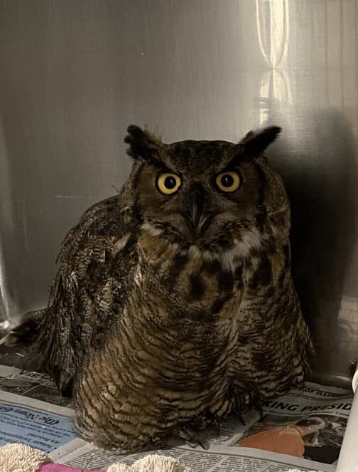
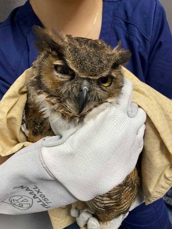
Bright and early on a Sunday morning, a good Samaritan made an over hour long drive to bring an injured Great Horned Owl, Bubo virginianus, to the WMC. The owl was reportedly found sitting on the highway the night before and was secured in a cardboard box until admission. On triage exam, the owl was found to be standing abnormally on the hock joints instead of perching on both feet but was otherwise bright, alert, and responsive. Upon further examination, it was found the abnormal stance was likely due to severe bruising, swelling, and abrasions to both feet and the left hock. The patient was reactive on examination of the left wing and dark purple bruising was noted at both elbows and the left carpal (wrist) joint. The owl’s eyelids were sunburnt and oddly, all the tail feathers were missing without any sign of trauma to the area. Ocular examination noted hyperpigmentation (darkening) on the ventral aspect of both eyes but no sign of active vitreal hemorrhage (bleeding in the eye) apparent. The owl was also quite well conditioned and was assigned a three out of five score, which is a good body condition score.
After examination, the patient was provided warm fluids, anti-inflammatory and pain relief medications to help with pain, and the injuries over both feet and hock were thoroughly cleaned. Though no fractures were palpated in the wings or feet, examiners wanted to ensure the patient was as comfortable as possible and did not suffer further injury in case a fracture was missed, so the left wing was secured in a figure 8 bandage and the patient’s enclosure was lined with thick fleece until a veterinarian was able to confirm the exam findings and radiographs could be completed. Bloodwork was completed and all findings were within normal limits.
Veterinary Exam, Radiographs, & Ongoing Treatment
Upon veterinary exam, much of the soft tissue damage to the feet had resolved, but bruising to the left carpi and both elbows were still present. Examination and radiographs confirmed there were no orthopedic abnormalities, despite the presumed traumatic event this owl had endured. Subcutaneous emphysema (air under the skin) was noted bilaterally on the flanks (sides) and ventrum (abdomen) but was not severe enough to impair respiratory function. This was one lucky owl! It was decided then to provide supportive care until the soft tissue damage resolved and this patient could be transferred to a long term facility to regrow the rectrices (tail feathers).
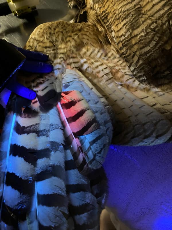
During this time, we were also able to age the owl using a Wood’s Lamp. Owl feathers, when they first grow in, contain porphyrins that fluoresce pink under UV light. As the feather is exposed to sunlight and ages, those porphyrins fade, leading to less pink fluorescence under UV light. Owls do not grow new feathers every year, except for their first set of feathers when they are nestlings. Given all feathers did not fluoresce the same hue, it can be inferred that the owl has lived long enough to go through at least one molt of adult feathers, indicating it is at least one year old, a true adult.
For supportive care, the patient continued anti-inflammatory and pain relief medications that were given in a food item during feeding. Treatment teams were instructed to monitor the patient’s mentation, ambulation, and for any injury to the carpal joints. We can also place padded bandages to the carpal area of the wing, called carpal bumpers, to give added protection to that area while the bird is housed in our hospital caging to help avoid further damage to the carpal joints. This was done in addition to fleece padding being added around the enclosure perimeters until the patient was approved to move out into our flight cage.
Reconditioning & Transfer to another Licensed Rehabilitator
When moved out to the flight cages, the patient was placed on the floor to observe flight capability. The owl was able to fly to the nearest perch with significant lift, so it was noted on track for reconditioning.
After ten days in care, this owl was successfully transferred to another licensed rehabilitator to continue flight reconditioning and give it time to grow back all its tail feathers. Prognosis for feather regrowth is fair to guarded given there may be damage to the underlying follicles when they are traumatically plucked, but only time will tell. We are hopeful that this individual will make a full recovery and be released back into the wild.
You’ve potentially found an injured wild animal and called the Wildlife Medical Clinic (WMC) to discuss what you should do. The volunteer at the WMC determines the animal should be brought in, but they tell you that they do not send volunteers out to pick up the animal, and that they can see the animal if you can bring it in. Oh no! You have never done this before! How can you safely capture and transport the animal??
Don’t worry, the WMC has advice on the dos and don’ts of transporting wildlife to the clinic. Remember, this is an incredibly stressful time for the animal, not only because of their injuries, but also because they are fearful of humans. It is important to know that proper capture and transportation is essential to the health of the animal. Stress from events prior to arriving at WMC can be very detrimental and could reduce the animal’s chances of being released! Do your best to follow these guidelines and to stay calm and quiet during this process.
Preparing for Capture
Gather capture materials before approaching the animal.
Having a towel, blanket, or other light-weight material to wrap around the animal is important for providing a barrier between you and the animal and can aid in controlling the animal.
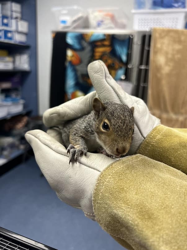
Thick work or gardening gloves are recommended to provide protection from possible bites and scratches.
An appropriately sized, secure carrier is important to have nearby to place the animal in after capture.
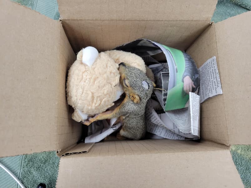
This can be a cardboard box, plastic bin with holes for ventilation, or a pet carrier. A container with an open top is not ideal because the animal could escape. The carrier should be lined with soft material that is not too stringy to avoid entanglement (T-shirt, towel, blanket).
Capture
Calmly approach the animal and completely cover it with the towel or blanket.
Carefully restrain the animal as you pick it up and place it into the box. Avoid handling the animal for longer than necessary.
Cautions
All wild animals have the potential to transmit disease to humans. Be particularly cautious with mammals, especially raccoons, coyotes and fox as they have the potential to transmit rabies if they are infected and you are bitten. This is why personal protective equipment (PPE), such as gloves and a towel, are very important. For mammal species that are carnivores, it may be advised to contact animal control for assistance with capture.
We unfortunately cannot accept any bats or skunks. If you find one of these species in distress, please do not touch the animal and call animal control for further assistance.
If capturing a raptor (hawks, eagles, owls, kestrels, falcons) be particularly cautious around their feet. While they can bite, they are more likely to talon you.
If capturing a heron or long beak bird, mind their beaks. They have long necks that can produce powerful strikes and a pointy beak at the end of that strike.
Avoid touching your face after handling the animal and its carrier. Washing your hands is a very important step to take after interacting with wildlife, even with gloves on! Disinfecting any items or surfaces that may have come in contact with the animal, the carrier, or unclean hands is a good idea as well.
Care
If you cannot come to the WMC immediately after capture, keep the captured animal in a dark, quiet, and warm place in the meantime to reduce their stress.
Do not attempt to feed or offer water to the animal. This can do more harm than good as they can aspirate or choke on what is offered. Additionally, the food may not be appropriate nutritionally as wildlife have very specific needs, especially young animals.
Transportation
Place the covered, secure carrier on the floor or seat of your car. Make sure the carrier will not rattle or slide around during transport.
Keep the car ride as quiet and smooth as possible. Noise will only stress the animal more during this time.
Do not let someone hold the animal during transportation as it is dangerous for you, them, and the animal. Bites and scratches could occur. Furthermore, the animal could escape their grasp and cause dangerous hazards while driving.
In Conclusion
Thank you for caring about wildlife and helping when you can! We hope these tips have relieved your concerns about bringing in a patient! If you have any reservations or need further guidance for an injured, ill, or orphaned animal you found, please reach out to us at 217-244-1195 and we will do our best to respond to you as soon as possible.
Check out the College of Veterinary Medicine’s blog post below. Dr. Lewis, Director of the Wildlife Medical Clinic, discusses highly pathogenic avian influenza (HPAI) and the risks it poses to you and your pets.
Wildlife Medical Clinic has successfully closed our 2024 chapter and opened our door to 2025. We are grateful for the support from our community, professional colleagues, fellow wildlife rehabilitators, student volunteers, and generous donors. With the collective force from everyone, we were able to provide the best possible care to the injured and orphaned wildlife we received. Without a doubt, our New Year resolution is to continue making a difference by providing excellent medical care for our current and future patients.
We wanted to share with you all the patients we cared for in 2024…
In total, our clinic saw a record-setting 2,278 patients come through our doors, comprising a remarkable 131 different species.
Of those 2,278 patients, we treated 7 amphibians, 46 reptiles, 768 birds, and 1,457 mammals.
Our most common species in the clinic was…
The Eastern Cottontail! They made up 580 of our patients last year.
The most common avian species seen…
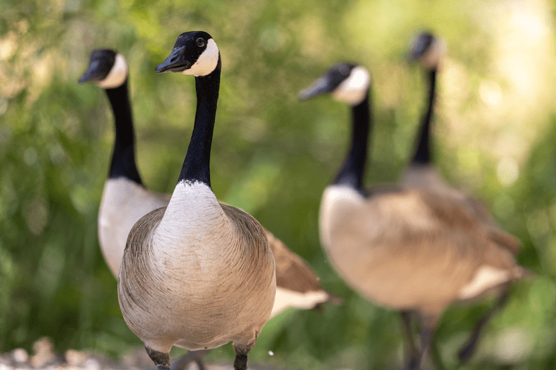
The Canada Goose, which enumerated 93 of our patients in 2024.
Our other commonly treated species included the Eastern Grey Squirrel, Virginia Opossum, Raccoon, American Robin, and Red-tailed Hawk.
Thank you all for your continued support of our clinic and the work that our wonderful team does every day to care for all these critters! We are excited about the new opportunities ahead and we will continue to provide care and services to our local wildlife with great passion.
Cheers to continuing to make a difference in the lives of Illinois wildlife in 2025!
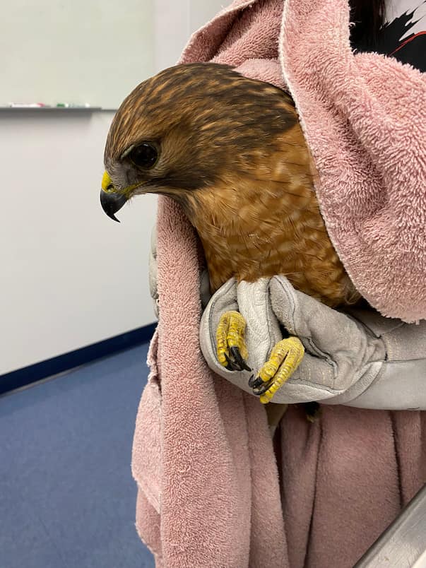
Juvenile red-tailed hawk 24-2042 arrived on September 14 with an ulnar (wing) fracture and ocular (eye) trauma. We provided pain relief medications, topical eye drops, and managed the ulnar fracture with a figure-8 bandage. Once healed, this patient was conditioned for release through a process called creancing.
What is Nutritional Secondary Hyperparathyroidism (NHSP)?
Nutritional secondary hyperparathyroidism (NSHP) is unfortunately a common type of metabolic bone disease that can occur in any animal but is most often seen in reptiles and amphibians. This condition most often occurs in captivity when an animal is not receiving the proper husbandry. NSHP can occur when the animal’s diet does not have enough calcium, or vitamin D3, or in the case of many reptiles when they do not receive enough UVB (ultraviolet B) light. Vitamin D3 is required by the body for calcium absorption, so as a result the parathyroid gland, which is found in the neck with the thyroid, begins to produce more parathyroid hormone (PTH). The primary function of the parathyroid gland is to tightly maintain calcium levels and keep them balanced. As a result of the decreased calcium and lack of vitamin D3 the secreted PTH stimulates osteoclasts in the bones, where most calcium is found, to begin resorbing the bones breaking them down causing the calcium to be released back into the bloodstream. Calcium is extremely important throughout the body where it is required for nerve transmission, muscle function, blood clotting and a multitude of other vital bodily functions. While the calcium needs to be in the blood it does so at the expense of the bones.
Why do we see NSHP in Wildlife?
When the bones are broken down to provide calcium it causes them to lose density making them more susceptible to injury. Additionally, there are developmental deficiencies associated with growth that can occur too. All of these make the lives of animals very difficult. NSHP is not as prevalent in the wild, because the animals tend to get enough sun and eat a proper diet. There are two main reasons we see this, first is when people have a pet and they don’t do proper research on housing and diet requirements and therefore don’t meet the needs of their pet. The second reason we see this is due to well-meaning individuals finding wildlife and then proceeding to care for them or keep them in captivity instead of bringing them to a licensed rehabilitator. Wildlife requires specialized diets and housing that are hard to meet in captivity and while people try their best, they often fail to meet the needs of these animals and don’t realize that for most wildlife, permits are needed to care for them, so keeping them in captivity is actually illegal if you don’t possess the proper permits. To further illustrate the effects of NSHP, we will review two cases we have recently seen in the Wildlife Medical Clinic.
Virginia Opossum Case
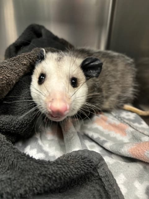
On September 9, 2024, we received a juvenile male Virginia opossum that a finder found in their yard struggling to walk. Upon exam he struggled to walk and lacked coordination in his back legs. Additionally, his face was swollen and asymmetrical. Due to the possible orthopedic issue and significance of ataxia we opted to take radiographs.
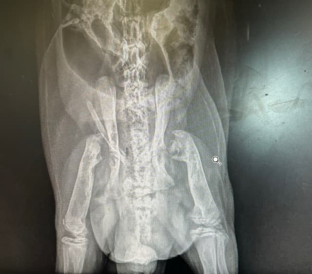
The radiographs showed that the opossum had a decrease in bone density, and severe malformations of the long bones primarily at the femoral head which is the part of the femur that connects to the hip. They were so degenerated that they appeared to be absent. All the joints appeared to be affected in some way, but that was the most notable location. These changes to the bones and the facial asymmetry led to our diagnosis of nutritional secondary hyperparathyroidism. Our belief is that he was kept as a pet for a significant amount of time when he was younger by someone who did not have the proper knowledge or credentials and then he was released. He also had a very high body condition score indicating he ate well, which would not have been possible on his own, in the wild, due to his musculoskeletal abnormalities. Even though he ate well, he did not receive the proper nutrients needed for bone formation and maintenance. Unfortunately, due to the severity of the malformations and the fact that multiple joints were affected the most compassionate care we could provide was humane euthanasia. He likely had arthritis, which is painful and overall had a poor prognosis for surviving in the wild as the damage done was irreversible.
Common Snapping Turtle Case
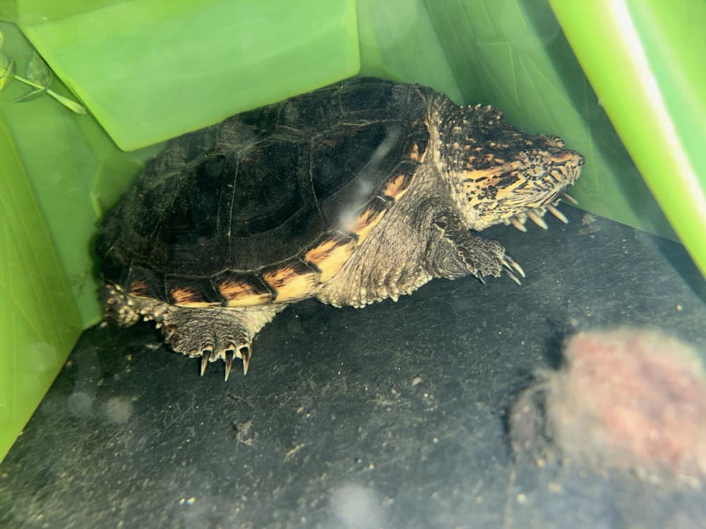
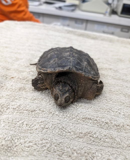
While the Virginia opossum did not have the outcome we hoped for, our next patient, a common snapping turtle, has a more positive outcome. On November 11, 2023, we received a juvenile male common snapping turtle after it was found abandoned in a vacated apartment where he was owned for around two years. He was found dry docked and severely dehydrated. Upon physical exam he was severely dehydrated, his shell appeared too small for his body and was soft and flat. Additionally, he had signs of stunted growth. All of this points to NSHP, primarily the softening of the shell and stunted growth.
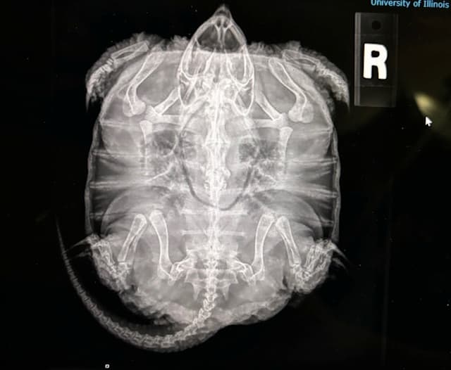
This was confirmed through radiographs, which showed decreased bone density and stunted growth Fortunately, unlike the opossum the damage was reversible. He was put on a proper diet along with consistent UVB and heat and now just over a year later he has grown a considerable amount and his body condition has greatly improved. While this is great news, he still is not releasable due to his friendly nature. Because of the time he spent in captivity, he is habituated to people. We have been working on finding him a permanent home and have received interest from an out of state nature center. However, if for some reason this home doesn’t work out, we will keep looking.
Lessons Learned from these Cases
These two cases illustrate the importance of nutrition and proper care for animals. If you want to own a pet, make sure you do the proper research and have everything set up and ready for them before you bring them home. Picking out a pet should not just be done on a whim. They are living creatures that require care and work. The snapping turtle shows the importance of doing research and being committed to owning a pet. Obviously, we don’t know the circumstances of his abandonment, but this is unfortunately an all too common occurrence when it comes to reptiles. Reptiles are unique in their dietary and husbandry needs, which need to be known by the owner, or else serious health issues like metabolic diseases can arise. The case of the Virginia opossum illustrates the importance of knowing who to contact, and only taking care of wildlife if you have the proper knowledge, credentials, and permits. The WMC is built on the idea of us working with the public to better the quality of life for wildlife. If you see an animal that you think needs help, don’t try to keep it and care for it on your own, because it can put you and the animal in danger. Contact a licensed rehabilitator or reach out to the Wildlife Medical Clinic and we can help provide guidance on what to do and who to contact. We hope these cases illustrated how important animal nutrition and husbandry is, why it is best to leave wildlife in the wild, and if help is needed to please contact a licensed rehabilitator.