Rabbit Fever, People, and Pets

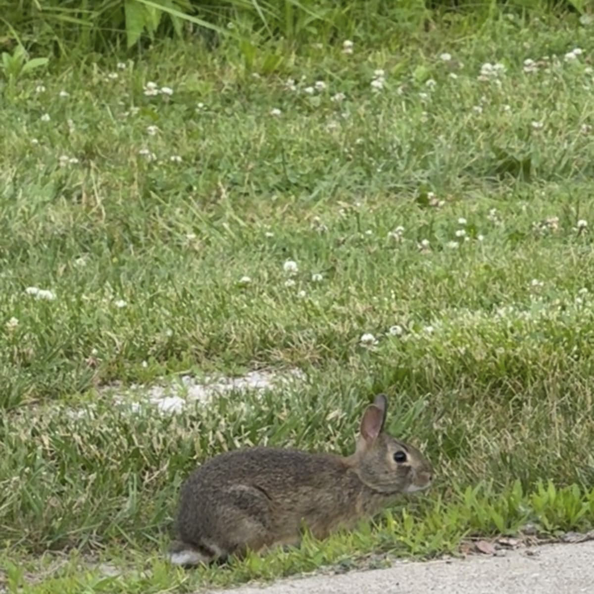
Earlier this year, the Champaign-Urbana Public Health District identified Tularemia, also known as Rabbit Fever, in several dead squirrels from the Champaign area. While Tularemia can affect people and animals, the lagomorph (rabbits and hares) and rodent populations are most susceptible to the disease. Both Eastern Cottontails (Sylvilagus floridanus) and Swamp Rabbits (Sylvilagus aquaticus) are native to the state of Illinois and could possibly act as sources of the disease.

What Is Rabbit Fever and How Does it Spread?

Rabbit Fever or Tularemia is a disease caused by Francisella tularensis, a gram-negative bacterium that is found in mud, dead animals, and water. Francisella tularensis can survive in the environment for about 4 months or up to three years in frozen meats, however, it is easily killed by disinfectants. There are two types of Francisella tularensis, Francisella tularensis biovar tularensis (Type A) and Francisella tularensis biovar holarctica (Type B). Type A is more harmful than Type B and is found in the North American region while Type B is less harmful and found in Europe and Asia regions.

Rabbit fever can be spread by direct contact with rabbits or other infected mammals via bite wounds, handling contaminated skin or paws, or in laboratory exposure. It can also spread via tick bites and inhalation of contaminated dust or soil. Additionally, it can spread through contaminated water and eating undercooked meat from an infected animal.

Signs and Symptoms in People and Pets

Tularemia can cause many different signs and symptoms depending on how the bacteria entered the body. The most common symptom in people is Ulceroglandular, which is caused by tick bites or after handling a contaminated animal. Ulceroglandular symptoms can be described as a formation of skin ulcers and swelling of lymph nodes in the arm pit or groin areas. Symptoms are also accompanied by a fever as high as 104°F.

Tularemia signs and symptoms in cats include fever, depression, appetite loss, and oral ulcers. Symptoms and signs in dogs include fever, loss of appetite, nasal and eye discharge, and abscess formation at the site of infection. Horses also exhibit signs of fever and depression, additionally they can show signs of trouble breathing, stiffness and swelling of the limb.

How to Prevent the Spread of Rabbit Fever

The spread of Rabbit Fever can be prevented in a multitude of ways, including the use of insect repellents, promptly removing ticks, wearing long pants and sleeves while outdoors, wearing PPE such as gloves and mask when mowing and working in your yard, and by avoiding drinking untreated surface water. Additionally, the spread of Rabbit Fever in pets can be prevented by limiting their exposure to wildlife, especially rabbits and rodents, and making sure that they are on Flea/Tick prevention year-round.
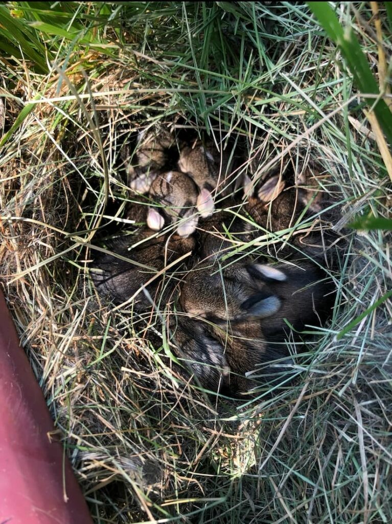
Many Eastern Cottontail neonates huddled together in their grassy nest.

Sources

Tularemia, U.S Centers for Disease and Control (2024, May 15th). Retrieved October 17, 2025, from https://www.cdc.gov/tularemia/about/index.html

Deaths Reported in Several Area Squirrels, Tularemia Identified, Champaign-Urbana Public Health District (2025, April 16th), Retrieved October 17, 2025, from https://www.c-uphd.org/press-release/2025/deaths-reported-in-several-area-squirrels-tularemia-identified.html

Tularemia, Cleveland Clinic (2022, August 25th) Retrieved October 17, 2025, from https://my.clevelandclinic.org/health/diseases/17775-tularemia

Tularemia in animals, AVMA, Retrieved October 17, 2025, from https://www.avma.org/resources-tools/animal-health-and-welfare/animal-health/tularemia-animals#:~:text=Enlarged%20liver,How%20is%20tularemia%20transmitted?

Written By: Irene J., Class of 2028
New Treatments of Chytridiomycosis

Chytridiomycosis is a fungal disease cause by Batrachochytrium dendrobatidis and Batrachochytriu salamadrivorans. This disease infects amphibians, effecting frogs the most. It has led to the decline and extinction of amphibian species across the globe. Scientists have been searching for ways to combat this disease. This article outlines two novel treatments of this disease.

Saunas for Frogs?

Although it sounds hard to believe, scientists in Australia have found that Hotspot Shelters, which are basically saunas for frogs, may be a new way to help combat Chytridiomycosis in wild frog populations.

Hotspot Shelters use the sun to generate heated shelters that use the fungus’ properties and frogs natural desire for heat to kill the fungus. B. dendrobatidis thrives in cooler weather and its virulence decreases in temperatures over 86 ℉ , which is the temperature inside a hotspot. In their study, scientists found that in both lab and semi-wild settings, frogs prefer to hang out in hotspots and that by hanging out in these hotspots for extended periods of time, they were able to clear Chytridiomycosis infections. Additionally, by clearing the infection, the frogs were able to develop immunity to B. dendrobatidis; thus, they are protected from future exposures to the fungus!

The benefits of hotspot shelters has been theorized by scientists for a while, but this is the first study to provide empirical evidence of their benefits. Hotspot shelters are relatively cheap and easy to make, which means they can be effective management tool of Chytridiomycosis in the wild! It is also theorized that hotspot shelters may be beneficial for other fungal diseases that effect wildlife such as White-Nose Syndrome in bats or snake fungal diseases!

Fighting Disease with more Disease?

Another potential treatment for Chytridiomycosis could come from secondary metabolitesof bacteria in the genus Xenorhabdus. Secondary metabolites are compounds produced by a living organism that don’t effect their growth, but offer some sort of advantage to the organism.

In this case, the secondary metabolites of interest are anti-fungals. The potential benefits of Xenorhabdus genus bacteria were first shown in 2023 and have been confirmed in a few other species since. All of these studies have found that in vitro, the metabolites from Xenorhabdus are able to completely halt the growth of B. dendrobatidis. When tested on living frogs infected with Chytridiomycosis, they found that treatment with Xenorhabdius metabolites was able to reduce the infection by almost 75%; thus, greatly increasing the survival of these animals. Additionally, no major side-effects were reported with this treatment. Although more research is needed, metabolites of Xenorhabdus species appear to be a potential treatment for Chytridiomycosis, especially in rehab settings where repeated treatments are possible!

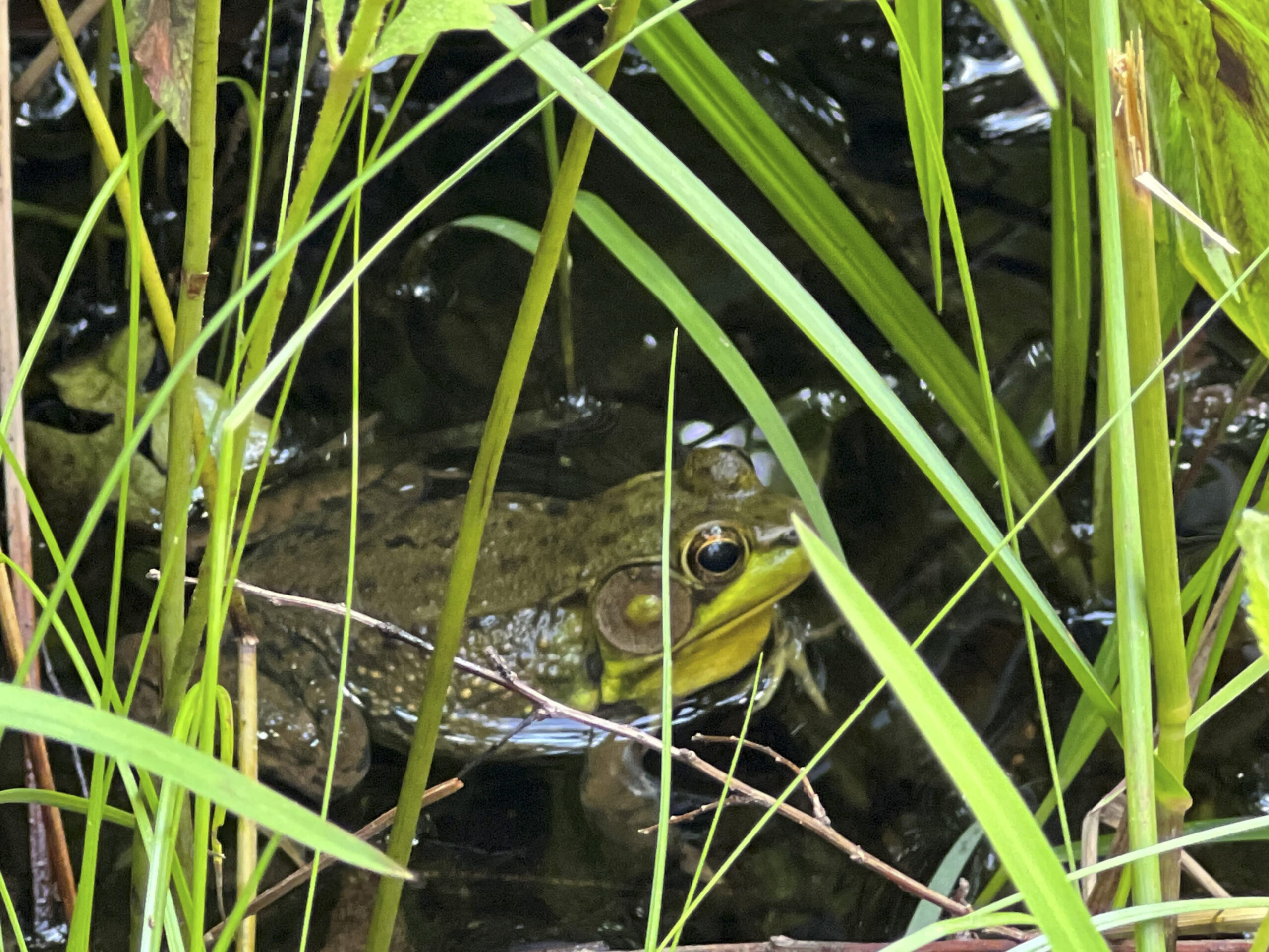
*The images in this article do not depict frogs infected with Chytridiomycosis. *

Sources

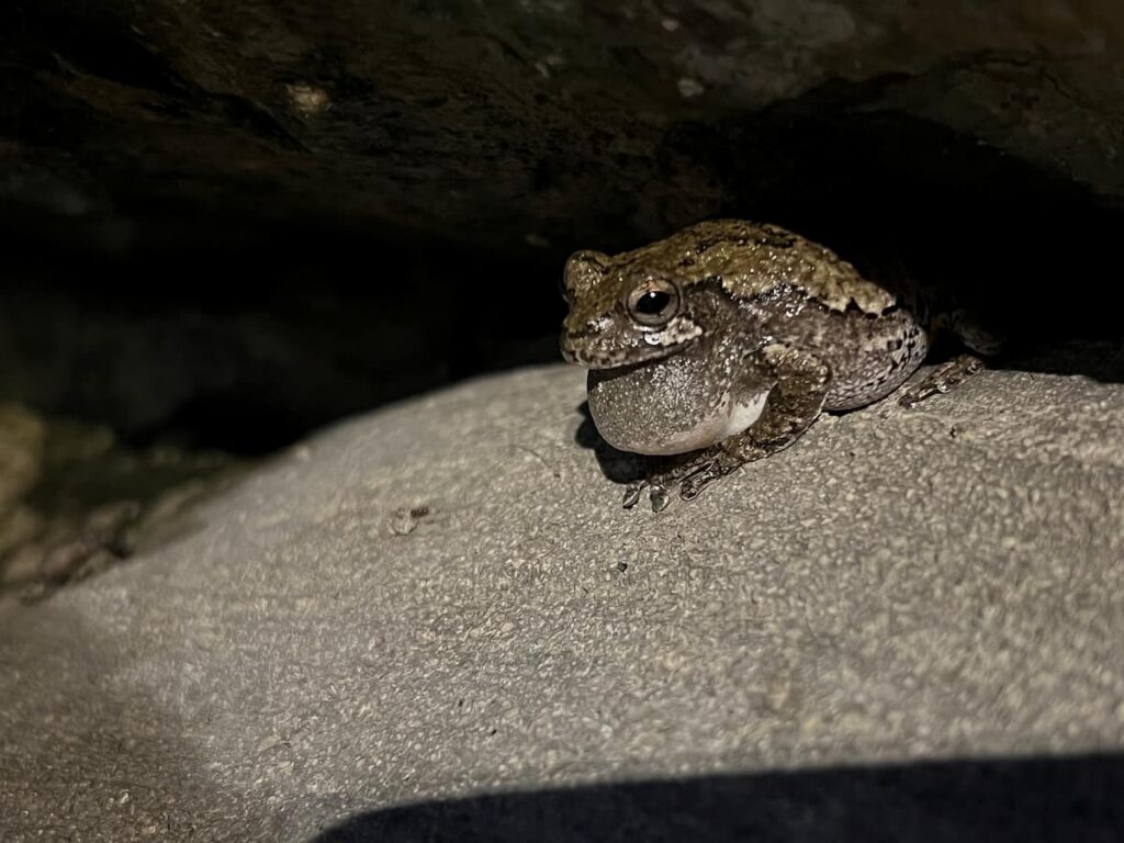
Gray tree frog calling on a rock.

Hotspot Study: Waddle, A.W., Clulow, S., Aquilina, A. et al. Hotspot shelters stimulate frog resistance to chytridiomycosis. Nature 631, 344–349 (2024).

Xenorhabdus Study: Ujszegi, J., Boros, Z., Fodor, A. et al. Metabolites of Xenorhabdus bacteria are potent candidates for mitigating amphibian chytridiomycosis. AMB Expr 13, 88 (2023).

Written By: Grace L., Class of 2028

Canine Distemper Virus: Not Just a Puppy Problem

What is Canine Distemper Virus (CDV)?

Canine Distemper Virus is a highly contagious and often fatal virus that can affect the respiratory, gastrointestinal, and nervous systems of animals. These locations make sense when considering these are all systems that contain rapidly-dividing cells, which the virus loves to replicate in. You may have also heard of CDV through your local veterinarian as protection against this virus is part of the core vaccine series recommended for every dog by the American Animal Hospital Association (AAHA). Among the consequences of infection, immune suppression is the #1 reason behind its mass mortality, as it leaves its host extremely susceptible to other diseases, particularly in young. Mortality rates for distemper can vary from 50% in adult dogs to even 80% in puppies, unvaccinated pets unfortunately being the most commonly affected. But despite its name, it is crucial to note that this virus does not limit itself to canines; CDV can infect a wide range of species!

Who else can be affected by CDV?

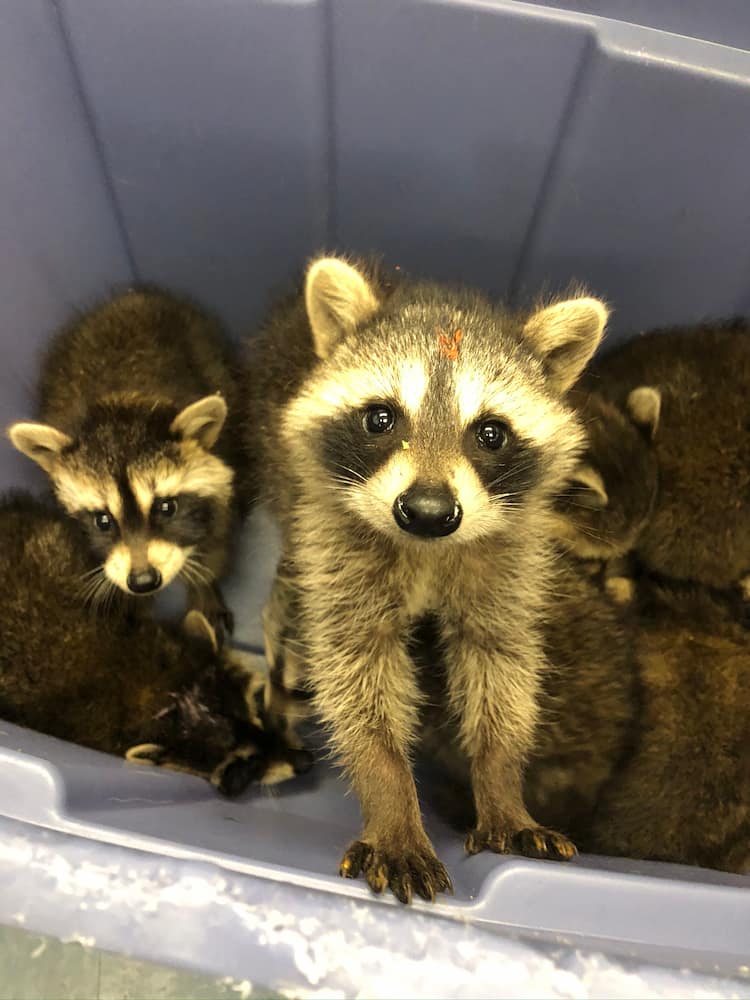
Tub of raccoons with one standing up.

While domestic dogs are the most well‑known hosts, Distemper can infect a variety of wildlife. Susceptible species include:

  • Raccoons
  • Foxes
  • Coyotes
  • Skunks
  • Ferrets — 99.9% mortality rate when unvaccinated
  • Domestic and large cats — less common
  • Some species of seals

This broad host range means that CDV can move between pets and wildlife with surprising ease. This is also how it creates a cycle of infection that is hard to break without implementing community-wide prevention efforts, which you may figure is no easy fit.

What makes CDV especially tricky is that animals can shed the virus for weeks, even when they look healthy. Just think about it, how many possibilities are there for any of these species to come into direct or indirect contact with our pets? A sick raccoon could wander through your backyard and spread the virus before it starts showing any symptoms. Your dog may also happen to interact with a seemingly healthy fox or other creature carrying the disease (or their biological by-products) during your monthly hike. It’s also easy to forget the route goes both ways. Our own pets, regardless of the source of infection, can also carry the disease onto others. The long list of hosts and possibilities of spread is one of the reasons CDV remains such an important One Health concern, as its impact can certainly extend past our homes and into the ecosystems around us.

What happens when an animal becomes infected?

CDV spreads through respiratory droplets (i.e. coughing or sneezing) and bodily fluids including saliva, urine, and feces. Once infected, there is a 10-14 day incubation period where the virus is settling in and replicating. An animal may then show initially mild and cold-like signs:

  • Fever
  • Watery eyes or nasal discharge
  • Tiredness
  • Decreased appetite

As the virus progresses, the symptoms can worsen as well:

  • Vomiting or diarrhea
  • Pneumonia (i.e. trouble breathing, persistent coughing, lethargy…)
  • Thickened paw pads (AKA, the classic “Hard pad disease” sign)

If the virus then reaches the nervous system, animals may develop neurologic signs like muscle twitching, seizures, or trouble walking (stumbling). Unfortunately, these are often permanent and can be fatal.

What you can do to help:

Vaccinate Your Pets:

Vaccination is the most powerful tool we have! The distemper vaccine has been proven safe, effective, and widely recommended for all dogs regardless of lifestyle. From an objective perspective, the benefits can outweigh the risks. It offers protection for your dog against the life‑threatening disease, prevents the virus from spreading to wildlife, plus helps keep local ecosystems balanced.

Feeders and Keeping Wildlife Wild:

Feeding wildlife or leaving food outdoors creates gathering spots where CDV can spread easily through increased close contact and shared surfaces. It is important to note that animals that become dependent on human‑provided food may also lose essential survival skills, making them more vulnerable over time. Allowing wildlife to find natural food sources is safest, but in the case you choose to feed certain species, consider using methods that limit cross‑species contact and keep feeders clean. Simple choices, like hanging bird feeders high enough to deter raccoons and foxes or using small openings that discourage squirrels, help reduce CDV spread and support healthier wildlife communities.

Report Sick Wildlife:

If you see a wild animal acting strangely (i.e. notice stumbling, twitching, or being friendlier than appropriate), contact your local wildlife rehabilitation center or animal control. These behaviors may signal CDV or another serious illness.

Stay Informed with Reliable Sources:

Because misinformation can lead to harm, it is crucial to aim for reliable, science‑based resources to stay up to date on related topics. Some examples of organizations that provide trustworthy information on disease outbreaks, vaccination guidelines, and wildlife health include:

  • American Veterinary Medical Association (AVMA)
  • Centers for Disease Control and Prevention (CDC)
  • World Organisation for Animal Health (WOAH)
  • Veterinary school and wildlife clinic websites
  • State public health or natural resources departments

As a final gentle reminder: Never hesitate to reach out! Your care makes a difference, plus people in this field of work are often happy to support you and grateful for your efforts.

Written By: Jennifer G., Class of 2026

Bats, Skunks, and Geese-Oh My!

Every day, the Wildlife Medical Clinic receives numerous calls from concerned community members about animals that appear sick or injured outside. Many of those people attempt to capture the injured wildlife, so they can be seen and treated at the Wildlife Medical Clinic. However, did you know that there are certain species you should use caution around and avoid, even if they appear to need help?

Bats and Skunks

Bats and skunks are two Illinois native species that the Wildlife Medical Clinic cannot receive. Why? These are two of the biggest vectors– animals that commonly transmit disease – of rabies virus. In fact, Illinois law makes it illegal for the wildlife clinic to take in sick or injured bats and skunks because of the high risk of rabies transmission, which would pose a major threat to the student volunteers, faculty, and community members that come into contact with these animals. Additionally, the rabies virus causes a multitude of different symptoms in wildlife, including profound depression, incoordination, and a lack of fear towards people — leading community members to stumble upon these animals more often. Our human instinct is to try and help the vulnerable critter, but Illinois state law and your friends at the Wildlife Medical Clinic urge you to stay far away. Wildlife, whether sick or not, often act unpredictably, and being bitten by a skunk or bat could potentially expose you to the rabies virus.

Waterfowl

Similarly, we encourage the public to keep their distance from geese and other waterfowl (ducks, swans, etc.) during the fall and winter months, due to a disease called Highly Pathogenic Avian Influenza (HPAI). Unlike the common flu people get, HPAI can cause severe neurological disease in our bird species, and has the potential to spread to humans and other domestic animals. Because of their migration patterns, geese and other water birds are currently the most frequent carriers of HPAI, which poses a public health risk if community members handle these birds directly – such as capturing them to bring into the clinic.

I found a bat, skunk, or goose that needs help. Now what?

The best thing to do is to call your local animal control facility. They have the proper equipment, training, and safety measures to be able to capture these animals, and avoid possible public endangerment. The Wildlife Medical Clinic will still accept and treat waterfowl like geese in the clinic, but only after they have tested negative for HPAI, which animal control can help facilitate by examining the birds for clinical signs before bringing them to the WMC. These protocols are in place not just to protect the public, but to keep our volunteers in the clinic safe as well.

After calling animal control, the best thing to do around sick or injured bats, skunks, and waterbirds is to keep as much distance as you possibly can. Do not attempt to handle these species directly under any circumstances, and avoid the painful (and expensive!) post-exposure-prophylaxis shots that must be given if you are bit or scratched by an animal that could carry rabies, or potential exposure to HPAI. We know it is tempting to want to help an animal in need, but we encourage you to leave it to the professionals when it comes to bats, skunks, and waterfowl, to keep everyone as safe as possible.

Written By: Kayla B., Class of 2027

Diagnostic Tools in Wildlife Medicine

Observation and Diagnostic Tools

When a good Samaritan brings in a Great horned owl that cannot fly or a lethargic Eastern box turtle with no apparent physical injuries to the Wildlife Medical Clinic, how do we determine what is going on with these patients? That’s where the problem-solving aspect of our work comes into play. In all fields of veterinary medicine, our patients cannot speak for themselves, and we must rely on clinical signs as well as the owner’s perspective of their pet’s behavior. However, wildlife patients are a little different in that they do not have owners who can speak for them. There are many different diagnostic tools we can utilize to aid in our understanding of what is going on with our patients, such as bloodwork and radiographs.

Bloodwork

In our bird and reptile species, bloodwork can be challenging to both perform and interpret due to a lack of reference intervals for every species and differences in cell morphology. Mammalian species have red blood cells that lack a nucleus. Birds and reptiles are special in that their red blood cells have a nucleus. This means that automated machines cannot accurately differentiate and count red blood cells and white blood cells. Anytime a blood sample is collected from a patient in the clinic, we want to manually determine a packed cell volume (PCV) and a total protein value (TP). We will also make a blood smear on a slide to later be evaluated under a microscope. In order to get the PCV and TP, we must first centrifuge the sample in a micro-hematocrit capillary tube which will separate the sample into 3 distinct layers. Red blood cells are at the bottom, white blood cells and platelets in the middle, and plasma at the top. We use a special card to aid in reading the PCV, or the percentage of red blood cells that are in the sample. Total protein is then measured by placing a drop of the plasma onto a tool called a refractometer. PCV and TP values can give us a clue on the hydration status of our patient as well as if they are anemic at that time. The blood smear mentioned before is used to evaluate cell presence and morphology. The presence of specific white blood cells can give us clues on if the patient has inflammation or even if there are microbial organisms within the cells. However, a challenge we often face in wildlife medicine is determining what values are normal as there often are no reference intervals available for many of the species we work with. Sometimes, we must make inferences based on what we already know about closely related species.

Radiographs

Another helpful tool we utilize often in all fields of veterinary medicine are radiographs. Any patients presenting with suspected orthopedic injuries will have images taken to be further assessed. We also take radiographs of birds who present with the inability to fly but have no obvious injuries to see if there are any fractures or dislocated joints. Radiographs can be helpful to determine the location and extent of these injuries so it can be decided whether or not the injury can be fixed. Imaging is also helpful for evaluating abnormalities in internal structures and can sometimes highlight the presence of metals that may have been ingested by the animal.

Conclusion

These diagnostic tests allow us to get one step closer to fixing our patients so they can be released back into the wild. Bloodwork and imaging are only two of the many tools available to us at the Wildlife Medical Clinic. If we suspect a specific disease, we can often run a specific test to either confirm our suspicions or rule that out. A closer look at what is happening inside of our patients, whether that is in their blood, skeletal system, or internal structures, can help guide us when creating treatment plans and determining a prognosis for our patients.

Written By: Grace M., Class of 2028

Grounded but not Defeated: An Owl’s Recovery Journey

Arrival and Initial Concerns

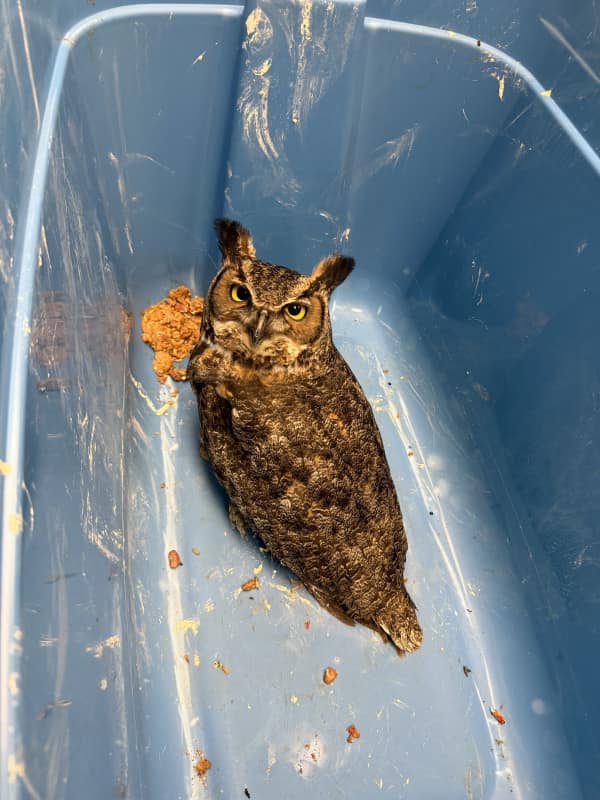
On August 31, 2025, the Wildlife Medical Clinic received an adult Great Horned Owl (Bubo virginianus) that had been found grounded and unable to fly. Upon intake, the owl was in thin body condition and demonstrated ocular abnormalities–most notably, aqueous flare in both eyes. This clinical sign indicates there is inflammation in the anterior chamber of the eye, raising concerns about systemic or infectious disease, or trauma to the eyes.

Despite its compromised condition, the owl was bright, alert, and responsive, but demonstrated an uncoordinated gait or ataxia. The combination of neurological signs, ocular inflammation, and poor body condition led the veterinary team to suspect West Nile Virus (WNV), a mosquito-borne disease well-documented in raptors such as Great Horned Owls.

Treatment Plan

The owl’s treatment plan was designed to address both the suspected viral infection and supportive care needs. It was started on:

  • Meloxicam: for pain relief and inflammation control.
  • Itraconazole: an antifungal agent, given prophylactically due to concern for secondary opportunistic fungal infections, as WNV is known to cause severe immunocompromise.
  • Excede: a long-acting antibiotic to treat opportunistic bacterial infections.
  • Ketorolac ophthalmic drops: to treat the inflammation within the eye.

Nutritional support was equally important. The owl was transitioned onto a diet of mice, often prepared as medicated feed items, which allowed medications to be delivered orally with minimal stress.

Progress in Care

Over the following weeks, the owl demonstrated steady improvements. The aqueous flare observed at intake resolved with treatment, and its body condition score increased as it began eating reliably. However, neurologic deficits–particularly ataxia–remained a significant concern. Veterinary student volunteers frequently documented episodes where the owl was able to perch normally but showed uncoordinated movements during flight attempts. On occasion, the owl even collided with enclosure structures when startled, reinforcing suspicions of WNV-induced neurologic impairment.

Despite these challenges, the owl displayed normal raptor behaviors: defensive posturing, hissing, and clacking at volunteers entering the flight cage. These responses indicated appropriate wariness of humans and reduced concern about imprinting.

Rehabilitation Transfer

By late September, the owl’s ocular inflammation had resolved, and it was maintaining weight well on a consistent diet. Its mentation improved, but residual neurologic deficits persisted. The Wildlife Medical Clinic does not have large enough enclosures to fully assess flight strength and coordination in recovering raptors, so plans were made to transfer the owl to the Illinois Raptor Center (IRC).

At the IRC, the owl would have access to larger flight enclosures, allowing for more thorough evaluation of its ability to hunt and fly effectively before considering release back into the wild. The transfer would also minimize unnecessary stress from prolonged handling in the hospital setting.

Conclusion

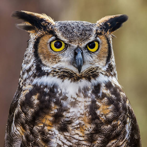
Close up of a Great Horned Owl.

This case highlights the complexities of managing West Nile Virus in raptors. While supportive care and anti-inflammatory treatment can help stabilize patients, neurologic deficits often linger and may impact long-term survival in the wild. For this Great Horned Owl, the combination of diligent medical care, nutritional support, and eventual transfer to a specialized rehabilitation facility offered the best chance of recovery and, hopefully, release.

West Nile Virus continues to pose challenges in wildlife medicine, reminding us of the interconnectedness between human, animal, and environmental health. By monitoring and treating these cases, clinics like the Wildlife Medical Clinic contribute not only to individual patient outcomes but also to broader understanding of wildlife disease dynamics.

Written by: Becca C., Class of 2028

References:

  • Nemeth, N. M., Beckett, S., Edwards, E., Klenk, K., & Komar, N. (2007). Avian mortality surveillance for West Nile virus in Colorado. The American journal of tropical medicine and hygiene, 76(3), 431–437.
  • DeCarlo, C., Omar, A. H., Haroun, M. I., Bigler, L., Bin Rais, M. N., Abu, J., Omar, A. R., & Mohammed, H. O. (2017). Potential Reservoir and Associated Factors for West Nile Virus in Three Distinct Climatological Zones. Vector borne and zoonotic diseases (Larchmont, N.Y.), 17(10), 709–713.
  • Wünschmann, A., Shivers, J., Bender, J., Carroll, L., Fuller, S., Saggese, M., van Wettere, A., & Redig, P. (2004). Pathologic findings in red-tailed hawks (Buteo jamaicensis) and Cooper’s hawks (Accipiter cooper) naturally infected with West Nile virus. Avian diseases, 48(3), 570–580

White-Nose Syndrome in Bats

Bats of Illinois

Illinois is home to a confirmed 13 different bat species who are all insectivores. Despite their feared reputation for carrying diseases such as rabies and feeding on blood, these bats are crucial ecologically and economically as they provide natural pest control, reducing insect pests for crops and saving farmers billions in pesticide costs. With insects in Illinois not being available year-round, different bat species must either hibernate or migrate to survive the winter. Bats that tend to hibernate in cooler areas, such as caves or abandoned mines, over the past decade have had their populations across North America decimated. Some common species in Illinois that are cool-cave hibernators are tri-colored bats, little brown bat, Indiana bat, and the northern long-eared bat which are now all endangered. The primary culprit of this phenomenon being White-Nose Syndrome. Other species that hibernate in warmer areas or have migratory patterns have been more resilient to white-nose syndrome.  

White-Nose Syndrome

In February 2006, a cave explorer in upstate New York discovered numerous bats coated with a white fungus. Researchers then launched an extensive investigation from Vermont to Virginia in the Winter of 2006-07 while they were hibernating to determine the extent of this previously unknown phenomenon. What they discovered was a fungus that was decimating entire bat populations, with mortality rates above 90%. Now close to two decades later it has spread to over 40 states and killing millions of bats. First spotted in Illinois in 2013, Pseudogymnoascus destructans is a fungus that thrives in cold and damp places that some bat species like to hibernate/congregate. It prefers to grow in temperature ranges of 39-59 Fahrenheit and will not grow above 68 Fahrenheit. As the fungus is not an airborne pathogen, the fungus is primarily spread from bat-to-bat contact. Due to this, bat colonies become rapidly infected during hibernation when they shut off their metabolism to save energy and are in close contact with each other.

Symptoms of White-Nose Syndrome

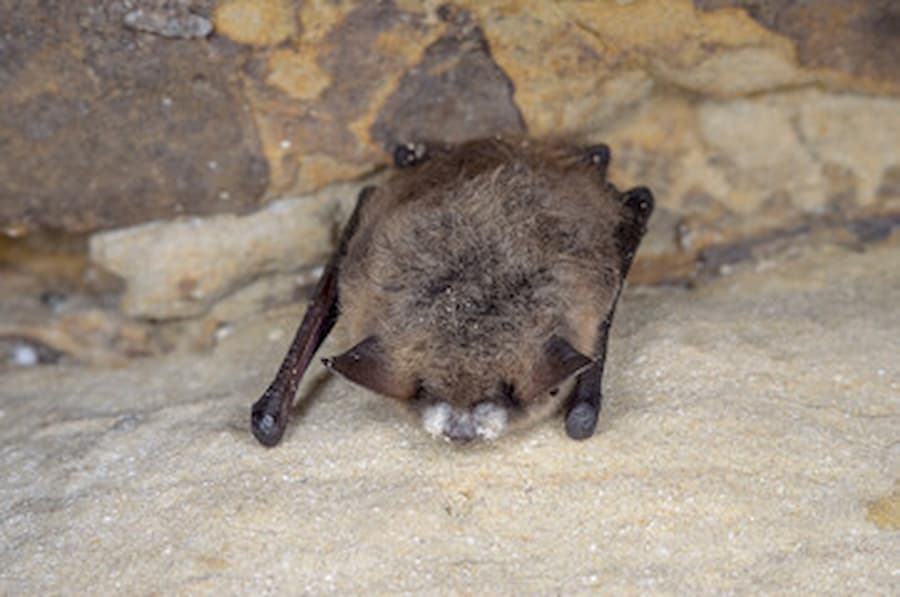
Bat with white muzzle indicating signs of white nose syndrome

Once infected, researchers have noted that the fungal infection causes hibernating bats to rouse, or awake from a state of torpor. Each time they rouse it causes them to use up their saved winter stores. Overtime, if the fungus causes the bat to rouse too often, it causes abnormal behavioral changes such as flying during the winter to try and feed instead of hibernating. Finding dead or dying bats on the ground in the winter is a strong indicator of white-nose syndrome. Other characteristic symptoms are the white growth of the fungus on the bats’ wings and muzzles.

Bats of all ages are susceptible to White-Nose syndrome and once infected there is currently no known effective treatment or cure. Researchers are currently developing an effective vaccine to protect colonies, UV-C light treated caves, probiotics to improve natural skin microbiome to slow fungal growth, use of volatile organic compounds (B-23), and temperature regulation are some preventative therapies being researched.

How to stop the spread of White-Nose Syndrome

Although the exact known cause of the spread of white-nose syndrome to North America is unknown, it is well hypothesized that human activity contributed to its spread from Europe. The fungus is naturally present in Europe and many of the bat species are well adapted as the mortality rates in Eurasian bat species are significantly lower. Additionally, bats are the only species known to date that can contract an infection from P. destructans and humans are unable to be infected. Due to this, researchers believe humans carry the fungus from cave to cave allowing a more rapid spread. Research has shown that the fungus can survive on clothing and surfaces for many years depending on conditions and advise anyone visiting a cave or mine to decontaminate afterwards. It is also recommended we do not wear similar equipment or clothing when visiting a different location with bats. Although bats that typically hibernate to survive the winter are mostly affected, it is important to remember that bats that migrate or hibernate in warmer conditions can also get infected or become carriers to spread the disease.

Written By: David K.

Works Cited

“Fighting White-Nose Syndrome in BATS Benefits Agriculture, Study Shows.” ACES Research News, CSU Press Release, 13 May 2022, https://aces.illinois.edu/news/fighting-white-nose-syndrome-bats-benefits-agriculture-study-shows.

Matteson, Mollie. “Devastating Bat-Killing Disease Reaches Illinois.” Center for Biological Diversity , 28 Feb. 2013, www.biologicaldiversity.org/news/press_releases/2013/white-nose-syndrome-02-28-2013.html.

“Nature Curiosity: What Is White-Nose Syndrome? | Forest Preserve District of Will County.” Forest Preserve District Will County , Reconnect with Nature , 4 Nov. 2024, www.reconnectwithnature.org/news-events/the-buzz/nature-curiosity-what-is-white-nose-syndrome/.

Vachula, Laura. “Preventing and Treating White-Nose Syndrome: U.S. Fish & Wildlife Service.” FWS.Gov, Wildlife Management, 28 Mar. 2023, www.fws.gov/story/preventing-and-treating-white-nose-syndrome.

“White-Nose Syndrome Faqs (U.S. National Park Service).” National Parks Service, U.S. Department of the Interior, 5 Nov. 2024, www.nps.gov/articles/white-nose-syndrome-faqs.htm.

“Wild about Illinois Bats!” Illinois Department of Natural Resources, Aug. 2019, https://dnr.illinois.gov/education/wildaboutpages/wildaboutwildmammals/wildaboutmammalsbats.html.

Turtles, Bunnies, and Cats, Oh My!

Why is it so important to keep our domestic animals inside and on a leash?

While pets love to have outside time and their pet owners love it too, it is important to be aware of your pet’s activities and supervise their time outdoors. They may come across wildlife that will not be as keen to meet them as they are to meet the wildlife! At the University of Illinois Wildlife Medical Clinic, we get many cases of cats and dogs interacting with wildlife that require our medical management. It is important to note that this can be risky both for our native wildlife and for our pets as well. The best way to limit interaction is to keep cats indoors or in a catio and walk dogs on a leash, even in the backyard.

What do we see most often at the clinic?

Throughout the year, we see Eastern Cottontail rabbits present to the clinic for different severities of wounds, including lacerations and broken bones often from being attacked by feral or outdoor domestic cats as well as domestic dogs. Treating patients who have been bitten or scratched by a cat can be challenging, and they must receive antibiotics promptly because cats harbor a bacterium called Pasteurella multocida. This specific bacterium can cause severe inflammation, redness, and even septicemia if it infects the bloodstream in rabbits, as well as our other mammalian and avian patients.

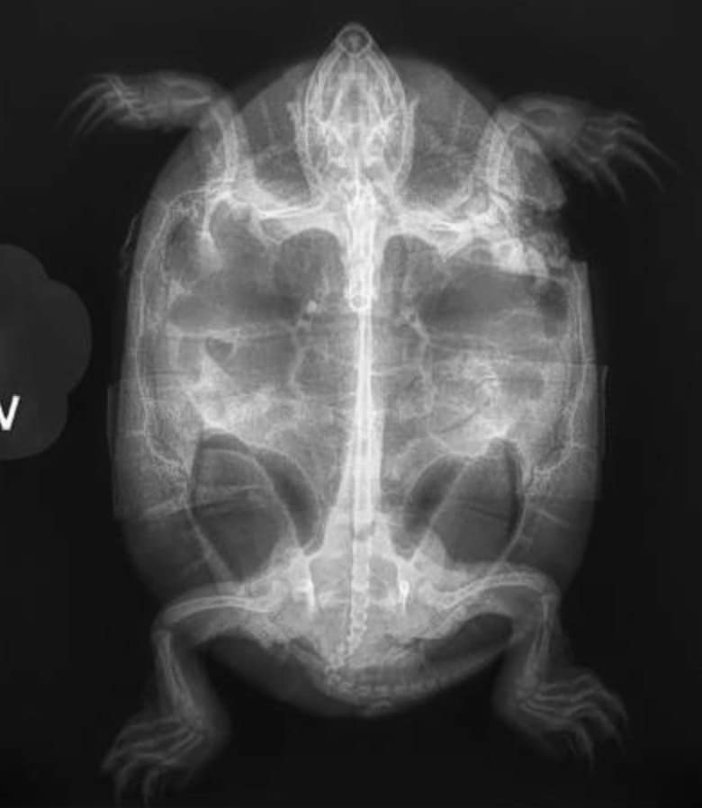
Painted Turtle x-ray

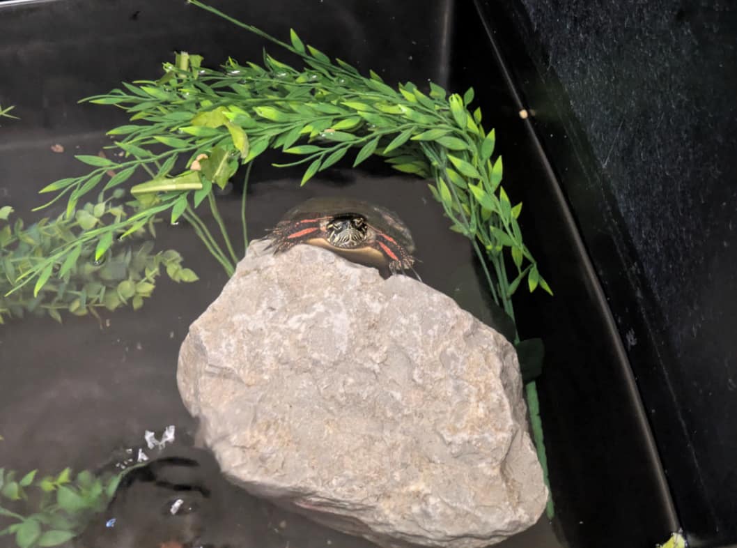
Along with rabbits, we also receive cases of turtles that sustain shell fractures when they are picked up by dogs. An example of this is Painted Turtle 25-966, who has been in our care since early June 2025, presenting to the clinic for several carapacial puncture wounds and shell fractures. The carapace is the upper portion of a turtle’s shell. After the initial intake exam, this patient was given the appropriate pain medication, antibiotics, and wound treatment. 25-996 will continue to heal in our care over winter, as it can take several months for these types of shell injuries to mend! He has been healing well, and the veterinary staff have seen positive improvements in his shell in the few months he has been with us.

How does all of this affect my pet?

While the Wildlife Medical Clinic does not treat domestic animals like our small animal medicine
colleagues do, we think it’s important to share how your furry family members can be adversely affected by wildlife interactions.

One type of infection that companion animals can pick up from wildlife is Salmonella. When dogs pick turtles up in their mouths, whether or not they think they look like their tennis ball or not, they can be susceptible to contracting an infection. Symptoms from this type of infection can include, but are not limited to, vomiting, fever, diarrhea, and lethargy.

Tularemia can also be a concern for outdoor cats and dogs that interact with wild rabbits in Illinois. This can be transmitted through direct contact with an infected rabbit or their body fluids, infected water sources, or blood sucking insects like ticks. Tularemia is a highly zoonotic disease (meaning it can spread between species and, in this case, to humans as well) and can cause symptoms such as fever, lethargy, poor appetite, enlarged lymph nodes, and abscesses. In humans, Tularemia can also present as ulcerating blisters that can spread the bacteria throughout the bloodstream.

What about birds? With the cooler season approaching, it is important to be aware of Highly Pathogenic Avian Influenza, or HPAI. While the name suggests, birds are the main susceptible species, cases have been seen in both cats and dogs, as well as people. One of the best ways to prevent your pets from contracting this influenza strain is by keeping pets indoors. Many cats and dogs outside may be attracted to birds, diseased or alive, or bird feces left behind, which can lead to the transmission of HPAI if touched or consumed. The main type of bird affected is waterfowl, but HPAI can be seen in any bird, including raptors and songbirds. Clinical signs in dogs and cats can consist of fever, lethargy, watery eyes, runny nose, difficulty breathing, and seizures.

If your pet interacts with any wildlife or is presenting with any symptoms, it is important to consult your primary veterinarian.

Helpful Links

https://vcahospitals.com/know-your-pet/tularemia-in-dogs

https://www.avma.org/resources-tools/animal-health-and-welfare/animal-health/avian-influenza/avian-influenza-companion-animals

Written by: Emily B., Class of 2028

White Nose Syndrome in Native Bats

Bats – Those that get a bad rap are under attack 

Bats are agile winged mammals that use echolocation to navigate at night as well as to hunt for prey. Bats usually live in caves in large colonies and vary in size and diet depending on the species. In recent years, bats have been under fire for being the source for several zoonotic diseases that can and have caused thousands of deaths in populations of other species. However, bats are an extremely important part within the ecosystem’s food web by clearing invasive and an overwhelming number of insects every night. For a place like Illinois, which thrives in the agricultural field, bats are a huge reason for healthy crop yields. Specifically in Illinois, there are five species of interest that are close to extinction due to the devastating effects of White Nose Syndrome (WNS). These species include the Little Brown Bat (myotis lucifugus), Indiana Bat (myotis sodalis), Gray Bat (myotis grisescens), Eastern Small-Footed Bat (Myotis leibii), and the Tricolored Bat (Perimyotis subflavus).

White Nose Syndrome

White Nose Syndrome (WNS) is a devastatingly infectious fungal disease stemming from the causative fungus, Pseudogymnoascus destructans (Pd). It is a disease that mostly affects hibernating bats, and it spreads rapidly from bat to bat. Pseudogymnoascus destructans (Pd) infects the skin on the ears, wings, and muzzle. This fungus erodes deep into the skin and disrupts the hibernation process of the bats causing physical and mental damage as they fight the infection while disrupted from their normal physiological sleeping cycles. Scientists found that the fungus is documented in the soil surrounding the caves providing easy access to the sleeping bats as well as their skin microbiome being a comparable community like the normal settling surface of P. destructans. Humans may also be potential carriers of P. destructans when they travel to national parks and caves, thus increasing the rate of infection.

WNS was first discovered in New York in the winter of 2006-2007. This disease has been devastating to many different bat species across North America, as the rise in total deaths caused by WNS has hit six million. In each bat population affected by WNS, it usually causes devastating effects of wiping out 90-100% of bat colonies. One of the main reasons that bat species have had debilitating effects from WNS is their low reproduction rate, as most bats usually have only one pup per year. Thus, if a bat population is almost completely wiped out from a WNS outbreak, it is difficult to recover the colony back to appropriate and healthy numbers.

How to Prevent the Spread and What are the Treatments

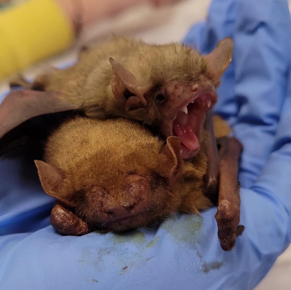
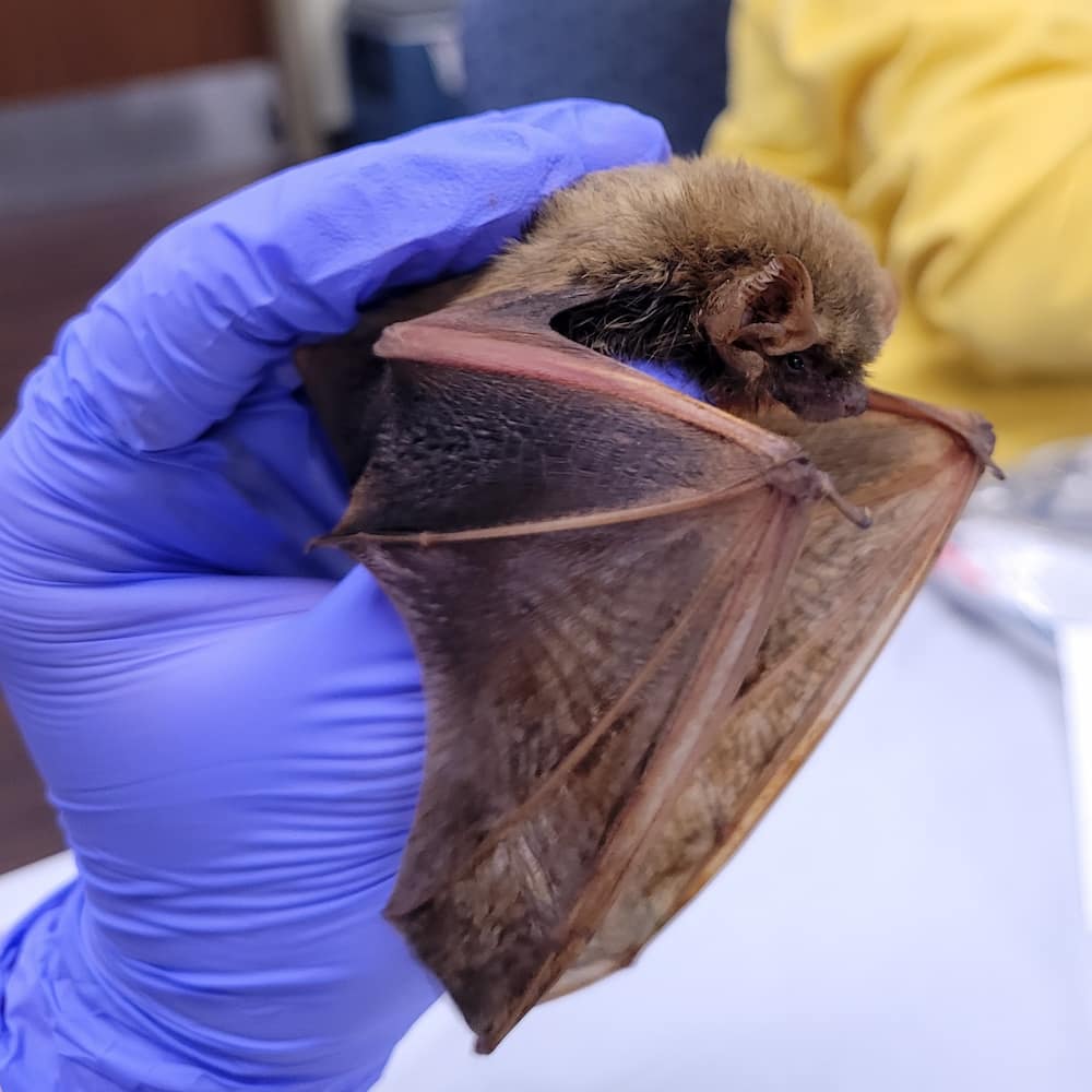
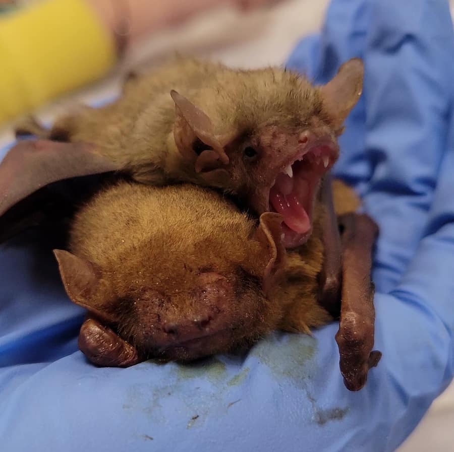
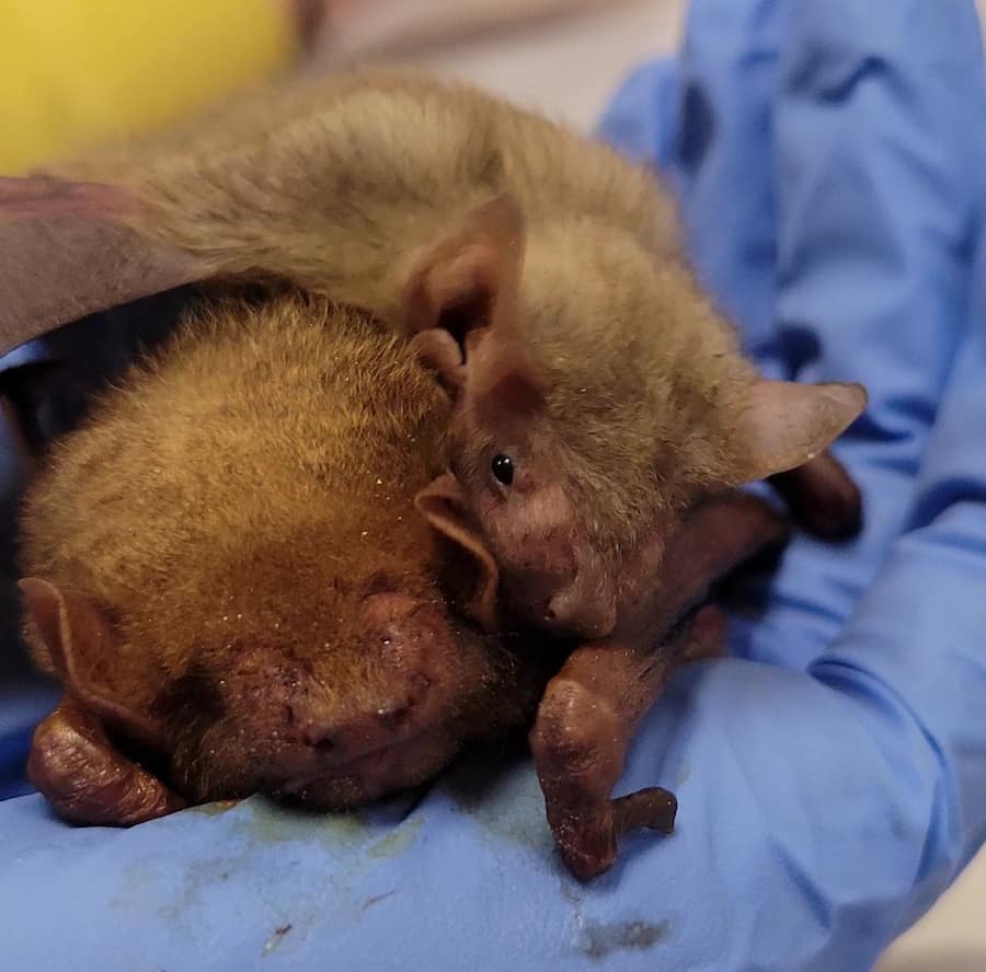
Two rehabilitation bat patients sitting on a gloved hand.

 As this disease is devastating many species across North America, the U.S. Government has been monitoring bat colonies to prevent continual spread and to help recover the already affected populations. The National Park Service has personnel monitoring bat populations, educating visitors about WNS and overall protecting their habitats. Decontamination processes, such as properly cleaning shoes and gear, are recommended as forms of prevention of further fungal spread as humans can be vehicles of transmission when entering multiple caves or mines.  As for treatment, scientists have produced a vaccine that uses a modified raccoon pox virus to express and attack fungal antigens as a protective measure. This is an oral vaccine that is given to bats in the fall or summer before hibernation processes begin. Scientists have noted that it takes about 3 years before the vaccine provides ample immunity in a colony; however, Idaho noticed a spread in 2021, after giving the vaccine the same year, they noticed in 2024 that they have no mortality in a specific cave bat colony. Since this vaccine is oral and requires human handling and capture, scientists are working on making aerosol sprays or larger scale vaccines and preventative treatments to minimize stress, human contact, and handling. While these diseases and animals can be scary to come into close contact with, if noticed while hiking or caving, it is important to identify the disease and report it to the proper local and federal authorities to better manage the spread of disease and limit devastating effects. 

Written by: Madison D., Class of 2028

Works Cited

Fighting white-nose syndrome in bats benefits agriculture, study shows | College of Agricultural, Consumer & Environmental Sciences | UIUC. (2022). Illinois.edu. https://aces.illinois.edu/news/fighting-white-nose-syndrome-bats-benefits-agriculture-study-shows

Powers, L., & Francis, B. (n.d.). Effects of white-nose syndrome on reproduction and survival in Illinois cave-hibernating bat species. Retrieved October 13, 2025, from https://dnr.illinois.gov/content/dam/soi/en/web/dnr/grants/documents/wpfgrantreports/20 14l16w.pdf 

Project T-78-R1. (2023). Illinois.gov. https://dnr.illinois.gov/conservation/iwap/white-nosesyndrome-and-il-bats.html

What species of bats are affected by White-nose Syndrome? | U.S. Geological Survey. (n.d.). Www.usgs.gov. https://www.usgs.gov/faqs/what-species-bats-are-affected-whitenose-syndrome  White-nose Syndrome FAQs (U.S. National Park Service). (n.d.). Www.nps.gov. https://www.nps.gov/articles/white-nose-syndrome-faqs.html

White-Nose Syndrome in North American Bats. (2025). USGS. https://www.usgs.gov/tools/white-nose-syndrome-north-american-bats

Will the killer White Nose Syndrome infect bats in Illinois? – News Bureau. (2025). Illinois.edu. https://news.illinois.edu/will-the-killer-white-nose-syndrome-infect-bats-in-illinois/

Wilson, D. (2020). bat | Description, Habitat, Diet, Classification, & Facts | Britannica. In Encyclopedia Britannica. https://www.britannica.com/animal/bat-mammal

Vachula, L. (2024). Preventing and treating white-nose syndrome | U.S. Fish & Wildlife Service. www.fws.gov. https://www.fws.gov/story/preventing-and-treating-white-nosesyndrome

Case Study: The Hoot on Owl Eyes

Intake and Treatment Plan

Earlier this year the Wildlife Medical Clinic received an Eastern Screech Owl, from one of our close partners the Illinois Raptor Center. Unfortunately, this little fella’ had an issue unlike most of our raptor patients.

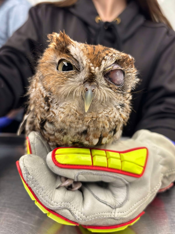
While you may be thinking, “Wow, I’m sure that owl would look super cute with an eye patch”, when we saw this guy’s eyes, we were concerned about potential disease processes that could be occurring to cause the eye to have this appearance. The grey-white color seen in this photo occurs due to cornea thickening and will often imply a permanent change causing vision loss in that eye. After a brief exam where we confirmed our suspicions but also verified that the other eye was functioning and healthy, our animal care team quickly set up a plan to perform an evisceration surgery to remove the non-functioning eye.

Can this owl be released with one eye?

Unlike most raptor species, owls can survive in the wild with only one eye. Many other raptor species (hawks, kestrels, eagles) are unable to be released if found with vision loss in one eye, due to the negative implications it can have on their ability to catch prey. Being diurnal species, or species that are active during the day, they primarily use their vision to hunt.

Owls, on the other hand, can hunt effectively even without perfect vision. Being a nocturnal species or species that are active at night, they primarily use their sense of hearing to hunt. Thus, allowing for their release with only one eye.

Post Surgery

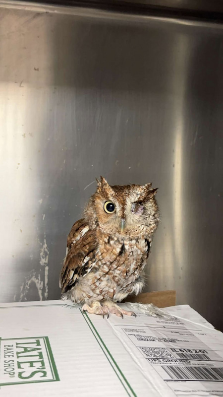
A one eyed Eastern Screech Owl in a hospital enclosure. It's injured eye was surgically removed.

This was our patient, following his evisceration surgery. He recovered great afterward and had no problem navigating his enclosure with only one eye! We were able to transfer this patient back to the Illinois Raptor Center where he continued his rehabilitation process with the end goal of eventually being released back into the wild!

When we say continued his rehabilitation process, this organization has larger enclosures that will allow them to flight test and condition this individual for release. They will ensure he can fly, find food on his own and exhibit species appropriate behaviors (confirm he is not imprinted or too friendly around people) before he is released back into the wild.

Written By: Katelyn D., Class of 2026

Wildlife Medical Clinic
John A. Coyne South Clinic
2100 South Goodwin Avenue
Urbana, IL 61801
217-244-1195