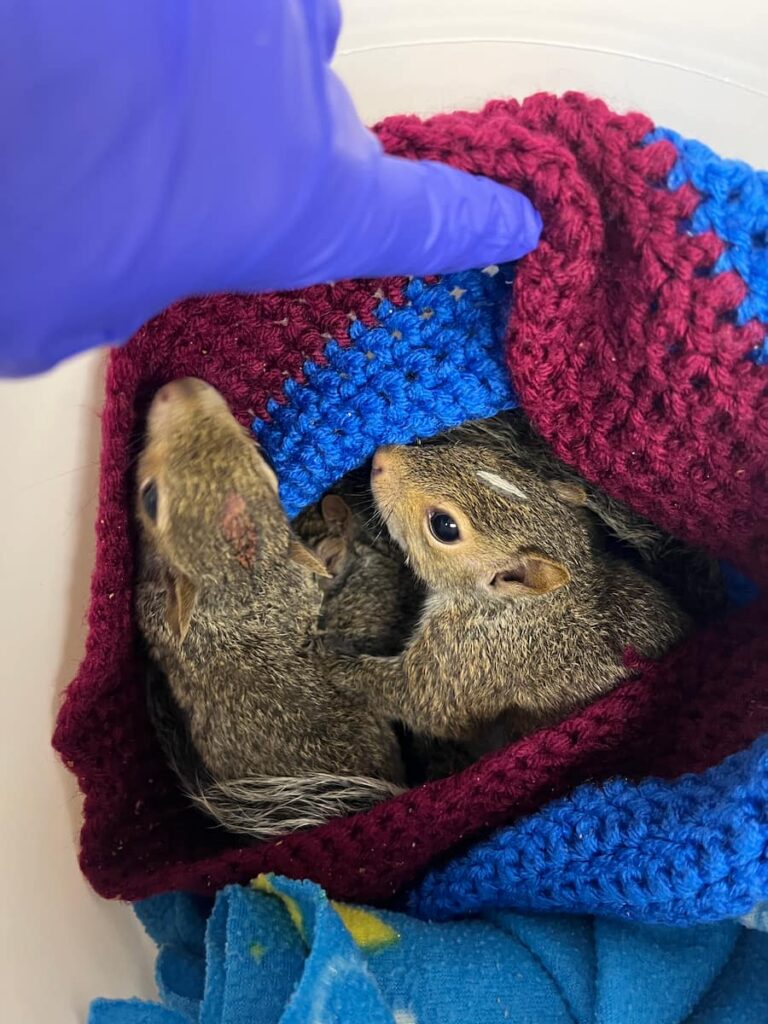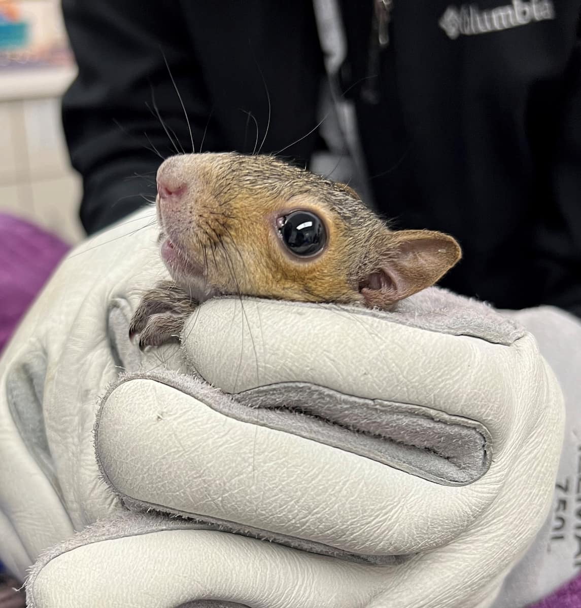
Eastern Gray Squirrel 25-1701

Initial Exam and Findings

Patient 25-1701 was an adult eastern gray squirrel that presented to the clinic on August 16th, for a possible head injury and abrasions. On examination, it was noticed that the patient had a broken incisor, but the extent of the damage was unknown. The patient’s wounds were lightly flushed, and the patient was started on anti-inflammatories and pain medication until radiographs could be performed to better assess its injuries.

Further Findings and Treatment Plan

On August 19th, the patient was sedated for a thorough exam and radiographs. On further examination under sedation, the patient was found to be missing a left upper incisor, with several healing abrasions on the rostral (front near the nose and mouth) aspect of the skull. Dental rads revealed a fracture of the remaining left incisor, but no evidence of a skull fracture was present. The patient was administered pain medication and was woken up from sedation. It was decided to allow the patient’s incisor to heal by second intent (or on its own) as the fracture did not extend to the skull or rear brachydont teeth and would theoretically heal over time. Due to the suspected head trauma which caused the broken incisor and abrasions, this patient was closely monitored and continued on a course of anti-inflammatories and pain medication through August 30th.

Ready for Release

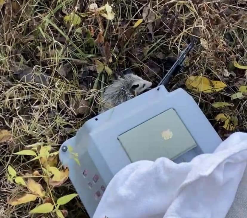
On September 5th, the patient underwent a recheck examination, which they were sedated for so that we could safely and clearly see the patient’s teeth. Healthy intact incisors, with normal length and growth were appreciated, with no oral swelling or malocclusion. At this time, the patient was off all medication, given a clean bill of health and was cleared for release. The following day, it was transport back to the area where it was initially found and released back into the wild.

Written By: London W., Class of 2028

A Troubled Turtle

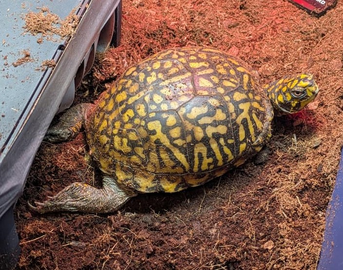
Eastern Box Turtle 25-611

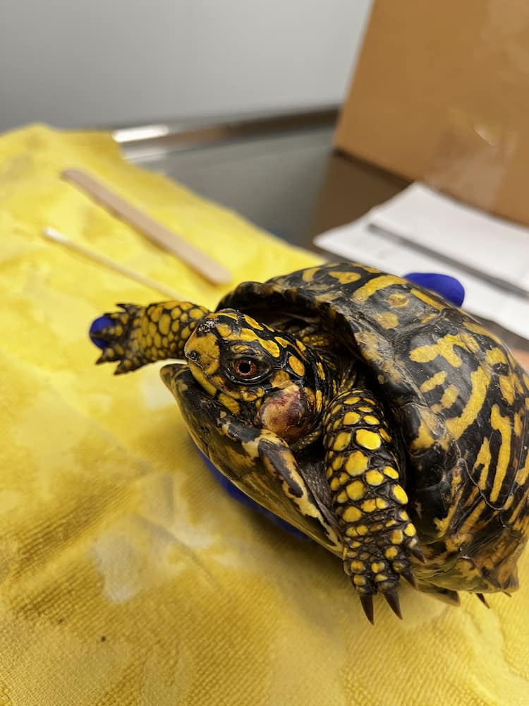
Meet 25-611, a male eastern box turtle. He came to us with an aural abscess–an issue commonly seen with turtles of multiple species, as well as some early warning signs of respiratory issues. Fun fact: the word “aural” means related to the ears, though it is pronounced almost identically to “oral”, which can cause confusion.

After spending a bit of time with us, it became obvious that this turtle was dealing with a serious respiratory infection. He was intermittently struggling to breathe, and symptoms were exacerbated when he was not at rest. Further evaluation was conducted with radiographs of his lungs, which showed signs of pneumonia.

Before we could fix his ear, we had to address his respiratory infection. He was started on a protocol of antibiotics and pain medication. After about two weeks on medication, we saw some improvement. However, discharge was seen coming from his left nare and eye, which indicated there was a blockage on that side called a rhinolith. Once he was stable enough for anesthesia, we were able to surgically remove the rhinolith along with the aural abscess.

So What’s Up with Aural Abscesses?

Aural abscesses in turtles happen when the turtle doesn’t get enough Vitamin A over a long period of time. Both wild and captive turtles get their Vitamin A from what they eat, but wild turtles don’t get vitamin supplements the way a pet might. When they develop a Vitamin A deficiency, their skin and mucus membranes near the ear start to change. The tissue overgrows and transforms into big clumps of dead cells that eventually become infected, interfere with the tympanum (the eardrum), and can even erode bone.

Once an aural abscess forms, it isn’t considered an emergency on its own, but it does require surgery to fix. Ongoing care is also needed to manage pain, treat any remaining infection, and possibly provide nutritional support.

Not Out of the Woods…

Luckily, 25-611’s aural abscess wasn’t particularly severe or complicated, and his surgery went well. Unfortunately, the surgery did not resolve all his issues. For a while, it looked like he was better. With his ear fixed and healing, his nose unplugged, and support from antibiotics, he gained weight and was breathing more normally.

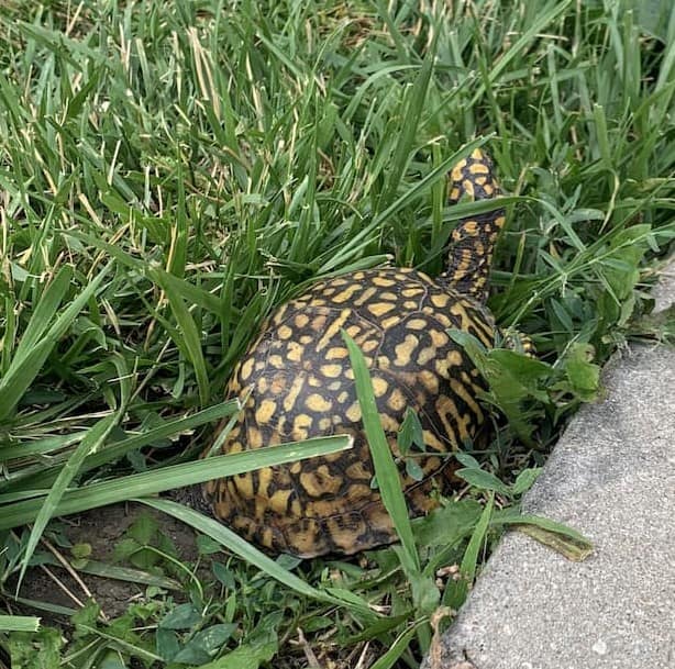
Eastern Box Turtle exploring outside next to a side walk.

In August, after two months, his lungs looked clear on imaging and his clinical signs had improved so much that he was taken off of medication entirely. Two weeks later, he was given one final check and appeared to have completely recovered from his pneumonia, so he was approved for release. Unfortunately, on the very same day that we planned to release him back to his home, his respiratory signs returned.

Strangely, though, they were a bit different this time. His x-ray images still showed that he was free of pneumonia, but his breathing would still intermittently be heavy, especially after exercise. We placed him back on two different antibiotics, and also pursued more aggressive diagnostics. We performed a lung wash, where a small amount of fluid is used to collect a liquid sample of whatever germs may be in the lungs. We also did a test for several important turtle pathogens. None of these provided any answers.

25-611 improved greatly and was off antibiotics, but release was not an option regardless as it was too late in the season and he would need to be overwintered and released in the spring when temperatures warmed up. We decided to proceed with high resolution CT imaging to confirm the pneumonia had cleared up and just to make sure there wasn’t anything that simple x-rays might have missed. Nothing new was found on the CT scan so we continued with the supportive care and closely monitored him over the winter months.

Approved for Release

Over the winter, 25-611 continued to improve and his appetite and weight increased and no further respiratory issues were noted. We just needed the temperatures to increase and stay consistent so 25-611 could be released back into the wild. After nearly a year in our care, patient 25-611 had a clean bill of health, was in great body condition, and the weather was finally suitable for release back into the wild!

Written By: Alex W., Class of 2028

Eastern Box Turtles, Frequent WMC Patients

The eastern box turtle is by far one of the most commonly received turtles that the Wildlife Medical Clinic (WMC) treats in its facility. Whether it be from an injury, illness, or anything in between, this turtle is one that the public finds often in our area and brings to the clinic when they are in need of help. While the eastern box turtle is named as a “turtle” they actually seemed to have missed the memo and instead spend a great deal of their time on land similar to a tortoise! They will enjoy a nice soak however, especially in the hotter summer season by burying themselves in some nice mud or by just going straight into shallow water.

Identifying Features of Eastern Box Turtles

Eastern box turtle with an aural abscess on left side of face.

The easiest way to know if you’re looking at an eastern box is by taking a look at the shell. They are known for their yellowish-orange speckled, high-domed carapace. The carapace is the fancy way of specifying the top part shell; the bottom part is called the plastron. Conveniently, their scaly, thick skin is actually a similar pattern too; it has an overall dark brown/black “background” with vibrant shades of yellow, orange, or red speckles. These turtles are relatively small, only reaching a size of about four to seven inches. Like many other turtles (and tortoises), eastern box turtles do not have teeth nor a bony jaw, alternatively, they have a jaw made up of a keratinized beak similar to that of a bird!

Common Causes for Intake

Eastern Box Turtle tucked in its shell. Can see wire on shell, repairing cracks.

There are many reasons someone might bring the eastern box turtle to our clinic. One reason could be that they accidentally hit one with their vehicle. Unfortunately, this is a very common occurrence as we continue to develop more land for agriculture and urbanization. So if you are driving through a road surrounded by more forested land, be aware of the road and drive the speed limit; if you are able to safely slow down, and/or go around the turtle this would be ideal for all parties involved. Oftentimes, female turtles are hit while searching for sites to lay their eggs. However, all sexes and ages are susceptible to vehicle collisions while migrating between food and water sources. Additionally, obstacles such as train track, curbs, and extensive debris pose challenges for the eastern box turtle trying to cross roads.

Another reason we may see them at the WMC is for trauma caused by domestic animals! So keeping a watchful eye and especially keeping your pets leashed is very important, not just for these turtles but other wildlife as well! Eastern box turtles may also be poisonous if other animals consume them since they are known for eating and snacking on poisonous mushrooms which then gets stored in their tissue as toxins. While their bodies can handle these toxins, the bodies of other animals may not fare as well.

How to Safely Move a Box Turtle

In the instance that you do come across an eastern box turtle that must absolutely be moved and/or brought in to be seen at the WMC, there are a few steps that you should keep in mind so as to avoid mishandling the turtle. If you just need to move them, move them in the direction they were travelling and move them just enough to get them out of harms way. They should not be moved from the area where they are found unless they are injured or ill. If you need to transport them to a licensed rehabilitator, please note the exact location the animal was found as that will be needed when it is time to release the animal back into the wild.

For handling, you can slowly and safely lift them using both hands, reaching between their front and back legs by placing your thumbs on the top shell (the carapace) and your fingers under the shell (on the plastron), almost like holding a hamburger. Avoid reaching in front of them or near the rear of them as to avoid having your fingers pinched when they close their shell. You should also avoid the front to reduce your chances of being bitten. Keep them relatively low to the ground as you are moving them in case they squirm and need to be set down or on the off chance that your grip is not tight enough and they fall, that way they are not falling very far. Alternatively, should you wish not to handle them with your hands, you can use a flat object (like a shovel or lid) to gently slide under them and transport them that way, still very low to the ground.

Next, place them in a well ventilated box or container that is large enough for them to comfortably turn around in, then make your way over to the WMC or another licensed rehabilitator as soon as possible. It is very important not to hold on to wildlife longer than absolutely necessary as this can cause stress and delay necessary treatment, hindering their chances of survival and release back into the wild.

Written By: Rachel L., Class of 2027

Bobcat Bulletin: A Wild Case Update

Bobcat (Lynx rufus)

American bobcats are a medium sized cat that can be up to two feet tall at the shoulders and weigh upwards of thirty pounds. They often have a mostly brown coat with black spots speckled throughout with a white stomach. They are found all throughout the United States and are very adaptable animals living in a variety of different habitat types.

A Unique Patient Not Often Seen in the Clinic

On December 3, 2025, the Wildlife Medical Clinic was presented with a sub-adult male bobcat that had been reported to have been hit with a car. Before being sedated on intake, he had a quiet mentation, didn’t want to move much in his carrier, and did not have much reaction to the people examining him.

Initial Assessment

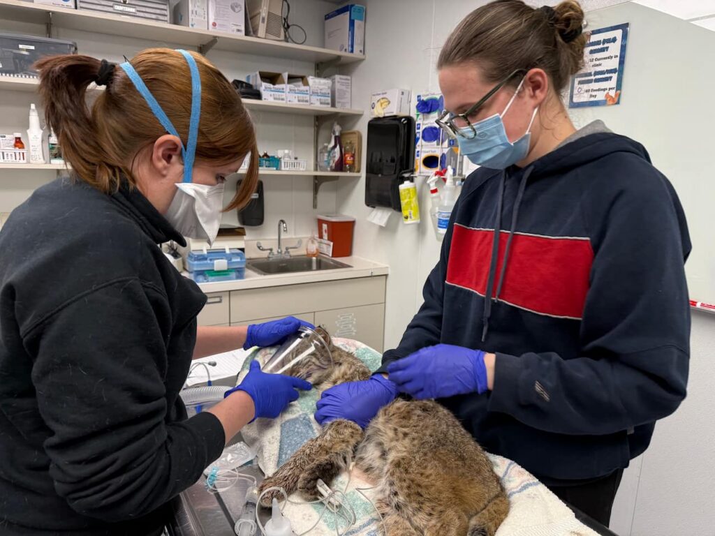
Triage exam on a bobcat. Two staff holding an oxygen mask over a sedated bobcats face.

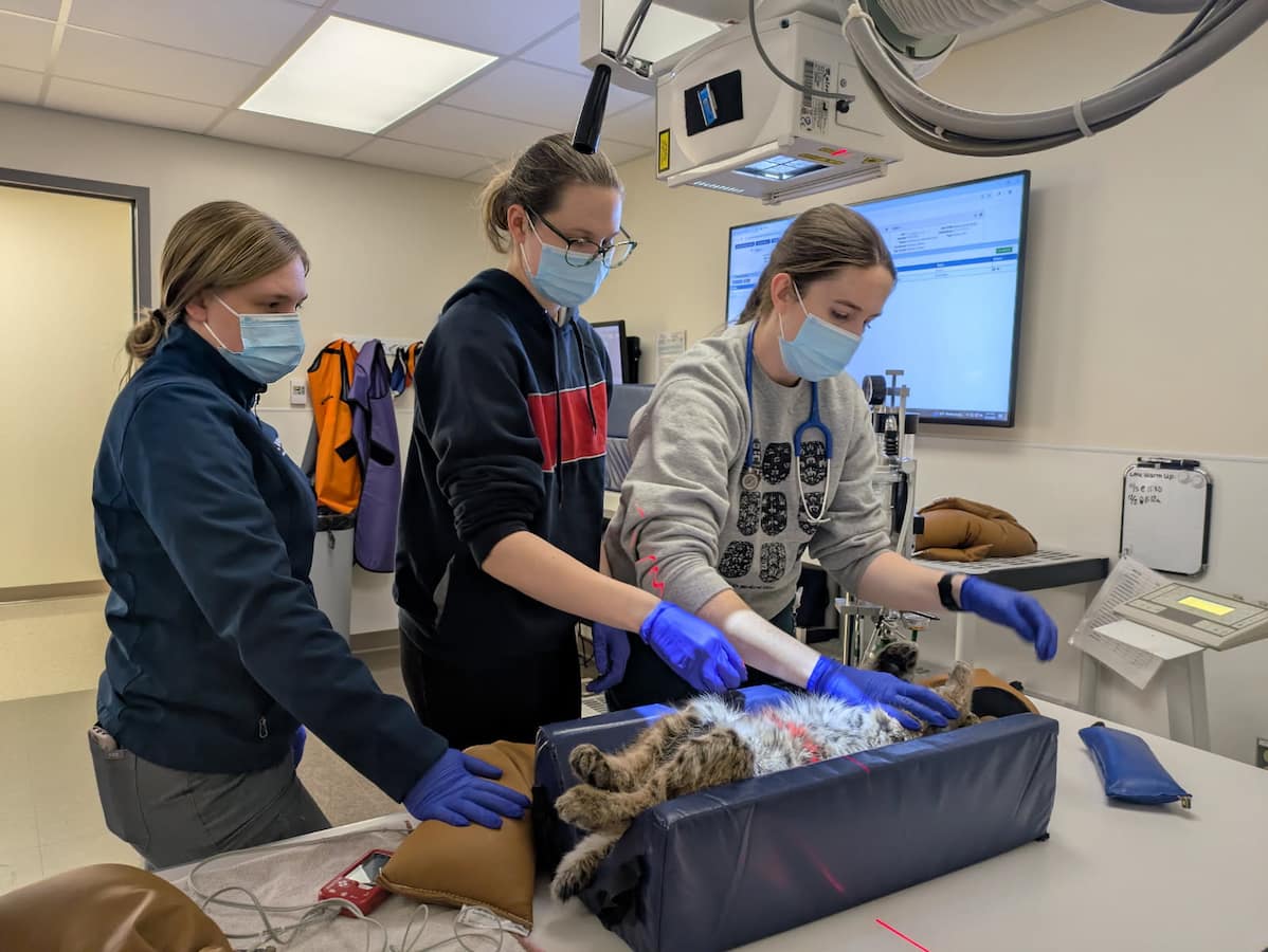
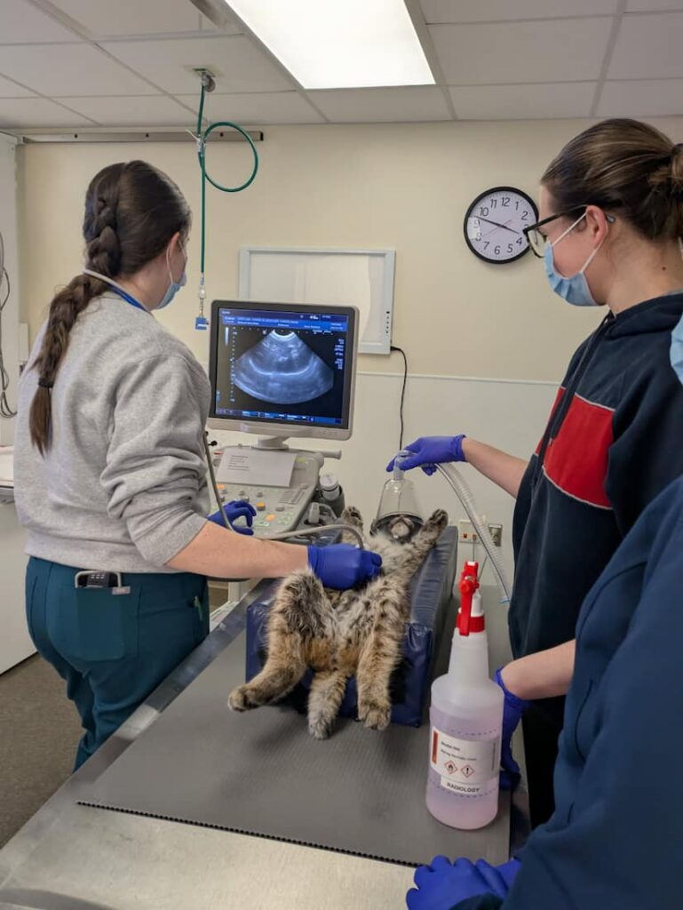
For his initial exam to ensure his safety as well as the safety of our staff he was fully sedated. This also allowed us to complete a thorough exam including: collecting a blood sample to run basic bloodwork and get a baseline on his overall health, x-rays, and an ultrasound. The blood work ended up showing mild signs of muscle injury, but no major signs of organ dysfunction. No broken bones or other major injuries were found on the x-rays. The point of care ultrasound was an abbreviated abdominal and thoracic ultrasound to look for areas of free fluid that could indicate bleeding into the body cavities; this ultrasound was also negative.

Our only concern at this point was the bobcat’s mentation. We started him on some pain medications, anti-inflammatory medications, and fluid therapy. The first round of fluids he received were given intravenous, or in the vein, in order to receive them quicker and help decrease any increased brain pressure. Additional fluids were given underneath the skin, subcutaneously, to give slower and ensure that he had adequate continued hydration. Now it was time to let the medications work and continue to monitor his symptoms.

Continued Care

Ultrasound of Bobcat.

However, over the next few days his mentation was still quiet and he wasn’t moving around the cage or hiding like we would expect, even with the continued pain and anti-inflammatory medications. We also had not seen him eating on his own. We decided to perform another sedated exam, and did another point of care ultrasound and reran the same bloodwork. The ultrasound was still negative and the bloodwork showed signs of improvement in the values indicating muscle injury. We decided to give another round of fluid therapy and also added some anti-nausea medication.

In the days after this exam the bobcat regained his appetite and began acting more appropriately by vocalizing when people were nearby the cage, and hiding from view when we were present. We continued to monitor over the next few days, and waited for the weather to be more appropriate for his release.

Final Assessment and Release

Our last step of the process was to perform an awake ambulation exam in one of our larger flight cages to ensure that he had full use of all his limbs and did not have any signs of limping or other injury. After being let out of the transport carrier he immediately ran and proceeded to climb up the wall with no issues or hesitation showing that he was back to normal. He received a clean bill of health from our staff veterinarian and was cleared for release. We were able to quickly secure him back in his carrier and he was immediately transported back to where he was found for release. Once we arrived and opened his carrier, it took a few seconds of figuring out where he was, but once he realized, he sprinted off into the woods and was gone in the blink of an eye.


Written by: Riley D., Class of 2027

A Stinky Situation

Case Intake

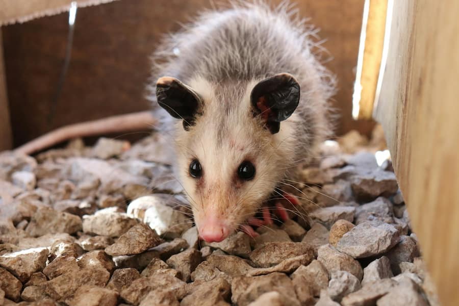
The Wildlife Medical Clinic has seen quite a few Virginia opossums this year and these are often a favorite species in the clinic due to their more agreeable attitudes and interesting anatomy. I had the opportunity to work closely with a few Virginia opossums this year, and DIVI 25-2152 was the first one where I was the veterinary student primarily in charge of his medical plan. DIVI 25-2152 was a male juvenile opossum who was brought into the clinic on October 10, 2025, after being found in someone’s septic tank. On his intake exam, we didn’t notice any apparent abnormalities or injuries, other than that he was a little bit dehydrated and that he had some gunk from the septic tank in his fur. He was displaying normal behaviors and proper defensive actions for his age. We gave him some subcutaneous (under the skin) fluids to help with his hydration status, cleaned up his fur, set him up in a comfortable cage, and offered food. As all seemed normal with him, we expected to just monitor for a couple days and then release after one last exam.

We ran bloodwork on the opossum a few days later to ensure that he was stable before releasing him. We tested his protein levels and red blood cell level (known as a PCV or packed cell volume), where we discovered that he did have slightly low protein levels and red blood cell levels, indicating that he was anemic. We additionally ran a test to see his blood lead level, which showed slightly elevated levels.

The Need for Lead Testing

Obtaining a blood lead level is a very important diagnostic test for the Virginia opossums that we receive at the clinic. Lead naturally occurs in the environment, but this environmental load has been greatly increased by human activity. Due to its previous use in paint, as well as its current use in batteries, shotgun pellets, bullets, and more, lead is pretty widespread within the environment. Virginia opossums are scavengers, which means that they do not directly hunt for their food and instead eat pretty much anything they can find, including dead animals. Due to this, it is highly likely for them to come into contact with and ingest lead, whether that is ingesting lead pieces accidentally, consuming roadkill with lead toxicosis, or consuming animals that contain lead pellets from hunting. With this in mind, almost all of the Virigina opossums that stay in the wildlife clinic will have their blood lead level tested. Many of them will have elevated levels slightly above zero, but they may or may not require treatment depending on how high that number is and how clinically affected they are.

Lead toxicity results in neurological problems in wildlife, causing things like head tremors, lethargy, paralysis, weakness, incoordination, seizures, and death in severe cases. With these issues, affected wildlife are generally less aware and are more likely to be injured, often being hit by cars or attacked by predators. These animals will often also lose weight due to their neurological condition and may end up fairly emaciated. The death of animals that have lead toxicosis tends to exacerbate the cycle, as scavengers, like birds of prey and opossums, will then potentially consume lead when feeding and become affected themselves.

Luckily, there are treatment options available for the opossums and other animals that present to the Wildlife Medical Clinic with lead toxicosis. Typically, they will receive chelation therapy, where medications like calcium EDTA or dimercaptosuccinic acid (DMSA) are used to bind to the lead in the body and help excrete it. After their initial chelation therapy has ended, we will collect blood again to check their blood lead level. Often, this will have decreased, and chelation therapy may either be discontinued or continued for another round depending on how high the animals level is and how clinically affected or symptomatic the animal is. Sometimes though, the blood lead level will increase. This doesn’t initially seem to make sense if treatments have been used to reduce the lead level, but this has to do with how lead is stored in the body. Some lead is present in the bloodstream and is targeted by our chelation agents, but a lot of the lead is stored in the bones of the animal, especially if they have had chronic lead exposure. This means that more lead will leak out into the bloodstream overtime and can increase the blood lead level from what it was originally despite treatment. In these cases, additional rounds of chelation therapy are conducted to continue to lower the animal’s overall lead level overtime, ideally getting their blood lead level down to a tolerable level for release.

Back to the Wild

Virginia Opossum being released back into the wild.

While this opossum’s blood lead level was higher than zero (12.4 μL/dL), we elected to not chelate this patient due to his normal attitude, behavior, and defensive actions, and his lack of apparent neurological signs. Additionally, he was overall pretty healthy and almost ready for release. To manage his anemia (low red blood cell levels) and hypoproteinemia (low blood protein levels), we kept this opossum in the clinic for about a week and offered him lots of food and fluids as he needed them. Since he didn’t appear to have any physical abnormalities or illnesses, it is likely that these low test values were due to him not having the best nutrition and likely being stuck in that septic tank for an extended period. A week later, he had gained over 300 grams, and his red blood cell and protein levels were within normal limits, so he was cleared for release by our veterinarians. He was released near where he was originally found, slowing venturing out into the forest. I was very glad to see this patient be released back into the wild and wish him the best!

Written By: Sarah F., Class of 2027

Case Study: The Fix on Fractures

The Eastern Box Turtle (Terrapene carolina carolina) is one of six subspecies of the Common Box Turtle and is found in the eastern United States. Eastern Box Turtles are mainly terrestrial turtles that live in areas of high vegetation and are close to small rivers or streams. Box turtles get their name from their hinged bottom shell, or plastron. This allows the turtle to completely close its shell around its body in dangerous or threatening situations. The plastron of an Eastern Box Turtle is dark brown, while their top shell, or carapace, can be bright yellow or orange. This coloring, and their four toes, is a helpful identifier of Eastern Box Turtles when they’re compared to other Box Turtle species. This coloring also allows the Eastern Box Turtle to camouflage among the damp earth, fallen leaves, and fallen vegetation in the highly moist and vegetative areas where they reside.

Intake

Box turtle with laceration under right rear leg.

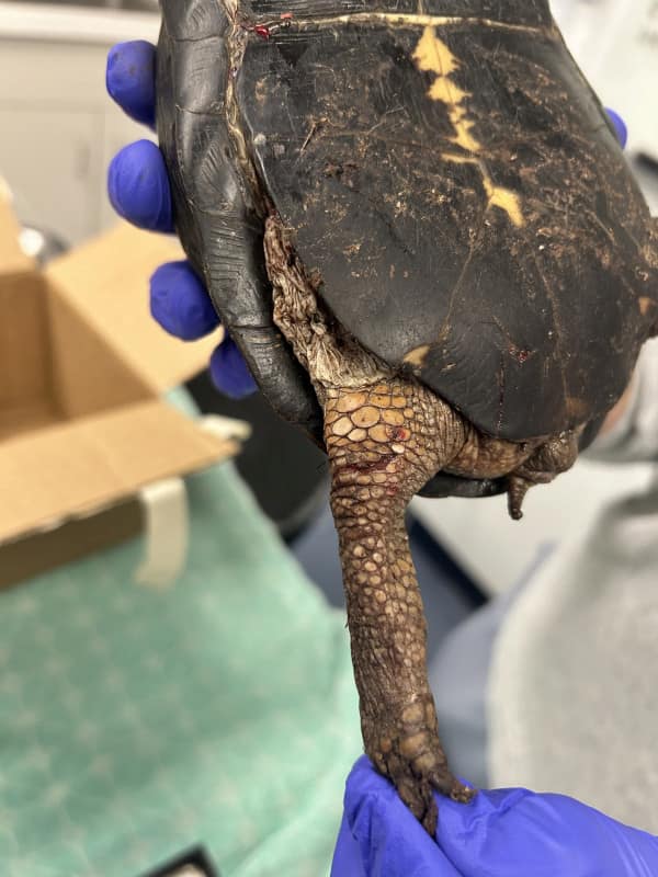
Eastern Box Turtle (TECA) 25-2096 is an adult female (as determined by her flat plastron and longer, thinner, curved rear claws) that presented to the Wildlife Medical Clinic on September 25 after being hit by a car. Upon intake, 2096 was alert, active, moderately dehydrated, and intent on wreaking havoc in the clinic, as she emerged out of her transport box mid-examination. 2096 had a superficial laceration across her right knee that did not appear infected and had a small abrasion at the midpoint of her right vertebral scute on her carapace.

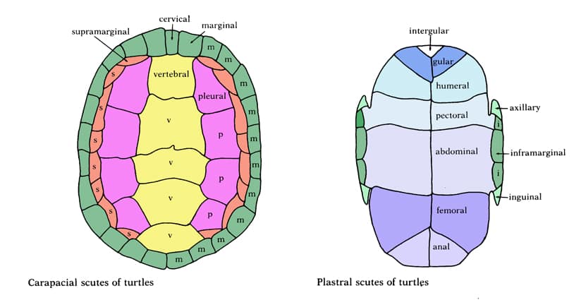
Drawing of a Carapace (top) vs Plastron (Bottom) for turtle shells.

On her plastron, which was the area of highest concern, there was a superficial, actively bleeding fracture at the level of her right marginal scute that, although not very wide, did extend all the way to her right femoral scute. She also had superficial fractures of her right anal scutes. Due to these injuries, initial treatments included subcutaneous (below the skin) fluids and an injection of hydromorphone, an opioid medication that is known to be helpful with alleviating pain from shell fractures. Vetericyn spray (an antimicrobial wound spray) was also sprayed over the laceration on the right knee to discourage infection and encourage healing.

Radiographs and Treatment

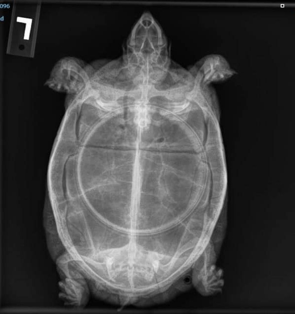
Not too long after her initial exam, radiographs were taken of TECA 25-2096 to determine the severity of her fractures. Three specific views were taken. There was a dorsoventral image, which looks down at 2096 from above, a left lateral image, taken from the right side of 2096, and a craniocaudal image, taken from in front of 2096 as if she was looking into a mirror. These three views are some of the most common views for turtles, allowing visualization of many important body parts and organs that cannot be visualized normally. The most beneficial view in 2096’s case was the dorsoventral view, which confirmed the fractures found on exam, and revealed two small fractures on the lateral most aspect of the right inguinal scute and a transverse fracture of the left inguinal scute. However, even with these numerous fractures, they were all minimally displaced and very superficial, meaning release was in the cards for 2096’s future. Post radiographs, the vetericyn spray was continued over the right hind limb laceration to encourage healing there, but the hydromorphone was switched to meloxicam (an anti-inflammatory) as the superficial fractures did not call for such strong pain medication.

Release

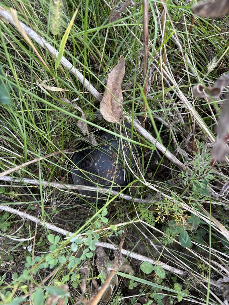
Box Turtle in tall grass being released back into the wild.

After nine days in the clinic, TECA 25-2096 was released into her habitat where she had been found in Tilton, Illinois. She was situated in an area of significant vegetation that bordered a small lake and was farther away from the road. Upon release, 2096 was eager to return to her home and quickly made her way into an area of high vegetation that allowed her to camouflage and hide within her new environment.

Works Cited

Eastern box turtle. Smithsonian’s National Zoo and Conservation Biology Institute. (2018, April 17). https://nationalzoo.si.edu/animals/eastern-box-turtle

Written By: Kat R., Class of 2028

Diagnostic Tools in Wildlife Medicine

Observation and Diagnostic Tools

When a good Samaritan brings in a Great horned owl that cannot fly or a lethargic Eastern box turtle with no apparent physical injuries to the Wildlife Medical Clinic, how do we determine what is going on with these patients? That’s where the problem-solving aspect of our work comes into play. In all fields of veterinary medicine, our patients cannot speak for themselves, and we must rely on clinical signs as well as the owner’s perspective of their pet’s behavior. However, wildlife patients are a little different in that they do not have owners who can speak for them. There are many different diagnostic tools we can utilize to aid in our understanding of what is going on with our patients, such as bloodwork and radiographs.

Bloodwork

In our bird and reptile species, bloodwork can be challenging to both perform and interpret due to a lack of reference intervals for every species and differences in cell morphology. Mammalian species have red blood cells that lack a nucleus. Birds and reptiles are special in that their red blood cells have a nucleus. This means that automated machines cannot accurately differentiate and count red blood cells and white blood cells. Anytime a blood sample is collected from a patient in the clinic, we want to manually determine a packed cell volume (PCV) and a total protein value (TP). We will also make a blood smear on a slide to later be evaluated under a microscope. In order to get the PCV and TP, we must first centrifuge the sample in a micro-hematocrit capillary tube which will separate the sample into 3 distinct layers. Red blood cells are at the bottom, white blood cells and platelets in the middle, and plasma at the top. We use a special card to aid in reading the PCV, or the percentage of red blood cells that are in the sample. Total protein is then measured by placing a drop of the plasma onto a tool called a refractometer. PCV and TP values can give us a clue on the hydration status of our patient as well as if they are anemic at that time. The blood smear mentioned before is used to evaluate cell presence and morphology. The presence of specific white blood cells can give us clues on if the patient has inflammation or even if there are microbial organisms within the cells. However, a challenge we often face in wildlife medicine is determining what values are normal as there often are no reference intervals available for many of the species we work with. Sometimes, we must make inferences based on what we already know about closely related species.

Radiographs

Another helpful tool we utilize often in all fields of veterinary medicine are radiographs. Any patients presenting with suspected orthopedic injuries will have images taken to be further assessed. We also take radiographs of birds who present with the inability to fly but have no obvious injuries to see if there are any fractures or dislocated joints. Radiographs can be helpful to determine the location and extent of these injuries so it can be decided whether or not the injury can be fixed. Imaging is also helpful for evaluating abnormalities in internal structures and can sometimes highlight the presence of metals that may have been ingested by the animal.

Conclusion

These diagnostic tests allow us to get one step closer to fixing our patients so they can be released back into the wild. Bloodwork and imaging are only two of the many tools available to us at the Wildlife Medical Clinic. If we suspect a specific disease, we can often run a specific test to either confirm our suspicions or rule that out. A closer look at what is happening inside of our patients, whether that is in their blood, skeletal system, or internal structures, can help guide us when creating treatment plans and determining a prognosis for our patients.

Written By: Grace M., Class of 2028

Grounded but not Defeated: An Owl’s Recovery Journey

Arrival and Initial Concerns

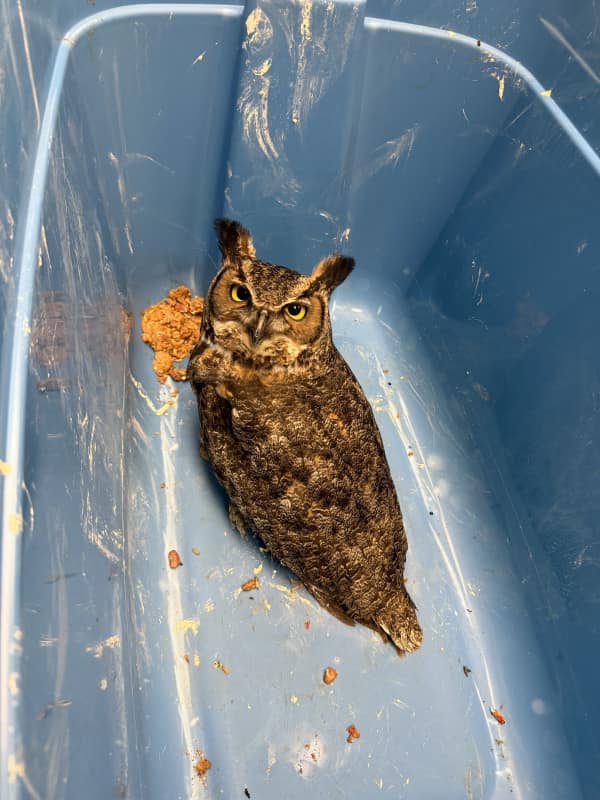
On August 31, 2025, the Wildlife Medical Clinic received an adult Great Horned Owl (Bubo virginianus) that had been found grounded and unable to fly. Upon intake, the owl was in thin body condition and demonstrated ocular abnormalities–most notably, aqueous flare in both eyes. This clinical sign indicates there is inflammation in the anterior chamber of the eye, raising concerns about systemic or infectious disease, or trauma to the eyes.

Despite its compromised condition, the owl was bright, alert, and responsive, but demonstrated an uncoordinated gait or ataxia. The combination of neurological signs, ocular inflammation, and poor body condition led the veterinary team to suspect West Nile Virus (WNV), a mosquito-borne disease well-documented in raptors such as Great Horned Owls.

Treatment Plan

The owl’s treatment plan was designed to address both the suspected viral infection and supportive care needs. It was started on:

  • Meloxicam: for pain relief and inflammation control.
  • Itraconazole: an antifungal agent, given prophylactically due to concern for secondary opportunistic fungal infections, as WNV is known to cause severe immunocompromise.
  • Excede: a long-acting antibiotic to treat opportunistic bacterial infections.
  • Ketorolac ophthalmic drops: to treat the inflammation within the eye.

Nutritional support was equally important. The owl was transitioned onto a diet of mice, often prepared as medicated feed items, which allowed medications to be delivered orally with minimal stress.

Progress in Care

Over the following weeks, the owl demonstrated steady improvements. The aqueous flare observed at intake resolved with treatment, and its body condition score increased as it began eating reliably. However, neurologic deficits–particularly ataxia–remained a significant concern. Veterinary student volunteers frequently documented episodes where the owl was able to perch normally but showed uncoordinated movements during flight attempts. On occasion, the owl even collided with enclosure structures when startled, reinforcing suspicions of WNV-induced neurologic impairment.

Despite these challenges, the owl displayed normal raptor behaviors: defensive posturing, hissing, and clacking at volunteers entering the flight cage. These responses indicated appropriate wariness of humans and reduced concern about imprinting.

Rehabilitation Transfer

By late September, the owl’s ocular inflammation had resolved, and it was maintaining weight well on a consistent diet. Its mentation improved, but residual neurologic deficits persisted. The Wildlife Medical Clinic does not have large enough enclosures to fully assess flight strength and coordination in recovering raptors, so plans were made to transfer the owl to the Illinois Raptor Center (IRC).

At the IRC, the owl would have access to larger flight enclosures, allowing for more thorough evaluation of its ability to hunt and fly effectively before considering release back into the wild. The transfer would also minimize unnecessary stress from prolonged handling in the hospital setting.

Conclusion

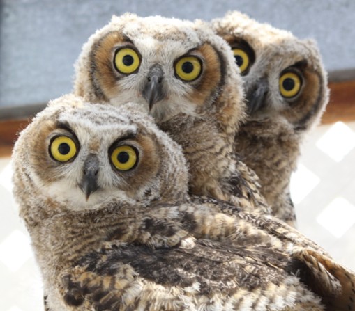
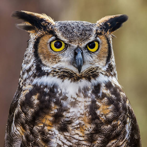
Close up of a Great Horned Owl.

This case highlights the complexities of managing West Nile Virus in raptors. While supportive care and anti-inflammatory treatment can help stabilize patients, neurologic deficits often linger and may impact long-term survival in the wild. For this Great Horned Owl, the combination of diligent medical care, nutritional support, and eventual transfer to a specialized rehabilitation facility offered the best chance of recovery and, hopefully, release.

West Nile Virus continues to pose challenges in wildlife medicine, reminding us of the interconnectedness between human, animal, and environmental health. By monitoring and treating these cases, clinics like the Wildlife Medical Clinic contribute not only to individual patient outcomes but also to broader understanding of wildlife disease dynamics.

Written by: Becca C., Class of 2028

References:

  • Nemeth, N. M., Beckett, S., Edwards, E., Klenk, K., & Komar, N. (2007). Avian mortality surveillance for West Nile virus in Colorado. The American journal of tropical medicine and hygiene, 76(3), 431–437.
  • DeCarlo, C., Omar, A. H., Haroun, M. I., Bigler, L., Bin Rais, M. N., Abu, J., Omar, A. R., & Mohammed, H. O. (2017). Potential Reservoir and Associated Factors for West Nile Virus in Three Distinct Climatological Zones. Vector borne and zoonotic diseases (Larchmont, N.Y.), 17(10), 709–713.
  • Wünschmann, A., Shivers, J., Bender, J., Carroll, L., Fuller, S., Saggese, M., van Wettere, A., & Redig, P. (2004). Pathologic findings in red-tailed hawks (Buteo jamaicensis) and Cooper’s hawks (Accipiter cooper) naturally infected with West Nile virus. Avian diseases, 48(3), 570–580

When a Shell, Isn’t Just a Shell

Intake

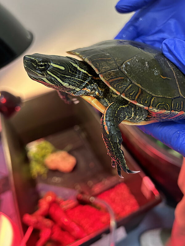
At the beginning of October, the Wildlife Medical Clinic welcomed a Painted Turtle brought in by a licensed wildlife rehabilitator from Illinois. Sadly, this patient had been the victim of a common — and often underestimated — threat to our native turtle populations: vehicle collision.

Many people are surprised to learn that when a turtle is hit by a car, the damage often goes far beyond what’s visible on the surface. A common misconception is that shell injuries aren’t serious. Unfortunately, this isn’t the case.

Shell Damage Is Skeletal Damage

A turtle’s shell isn’t just a protective outer layer — it’s a vital part of their skeletal system. Their ribs and spine are fused directly to the shell, which means that any cracks or fractures are often accompanied by internal trauma.

Our Painted Turtle patient arrived with multiple shell fractures, affecting the carapace (top shell), plastron (bottom shell), and bridge (the sides of the shell that connect top and bottom). These injuries alone were concerning — but we knew there could be more going on beneath the surface.

Imaging Reveals Deeper Injuries

To assess the extent of internal trauma, we performed radiographs (x-rays). Unfortunately, the images revealed fractures to multiple bones of the pectoral girdle of both forelimbs. The pectoral girdle are bones near the shoulder that are essential for forelimb function. These types of internal injuries significantly complicate recovery and reduce the likelihood of successful release.

How Did We Approach This Tough Situation?

Reptiles like turtles have slow metabolic rates, meaning healing takes much longer than in mammals — often over a year for serious injuries. Surgical repair of the bones of the pectoral girdle is not feasible in turtles, though minor injuries can heal with time and rest. In this patient’s case, its injuries were catastrophic, impacting both forelimbs, and unlikely to result in a functional outcome for either forelimb. This species of aquatic turtle requires function of at least 3 of 4 limbs for success in the wild post-release. Additionally, severe trauma to the pectoral girdle is often accompanied by extensive trauma to the lungs, the cranial aspect of which are adjacent to these bony structures.  While we evaluated options, we ensured the turtle had a quiet enclosure, pain management, and food.

A Difficult Final Decision

After much consideration, our veterinary team made the difficult decision to pursue humane euthanasia. While our clinic regularly treats turtles with shell and limb injuries — many of which go on to be successfully released — the severity of this patient’s internal injuries, paired with her lack of appetite and movement, made recovery highly unlikely.

Our goal is always to prioritize quality of life, and in this case, euthanasia was the most compassionate and medically appropriate choice.

What Can You Do?

During the fall, turtles often cross roads in search of overwintering sites. You
can help protect them by:

  • Slowing down and staying alert near wetlands, forests, or creeks
  • Contact a licensed wildlife rehabilitator if you find a turtle that appears injured — even if it seems like it’s okay. Prompt medical attention can make a life-saving difference.

Thank you for supporting our work and helping protect native wildlife through awareness, compassion, and action!

Written by: Natalie L., Class of 2028

Works Cited:

Hirasawa T., Nagashima H., Kuratani S. The endoskeletal origin of the turtle carapace. Nat Commun. 2013;4:2107. Retrieved October 17, 2025, from https://pmc.ncbi.nlm.nih.gov/articles/PMC3715867/

Jackson DC. Hibernating without oxygen: physiological adaptations of the painted turtle. J Physiol. 2002 Sep 15;543(Pt 3):731-7. Retrieved October 17,2025, from https://pmc.ncbi.nlm.nih.gov/articles/PMC2290531/

Virginia Zoo in Norfolk. Where did the turtles go for the winter? 2023 October 20. Retrieved October 17, 2025, from https://virginiazoo.org/where-did-the-turtles-go-for-the-winter

Ambassador Animals To The Rescue

On February 5, 2026, a juvenile Red-Tailed Hawk presented to the Wildlife Medical Clinic emaciated and with a depressed mentation, not able to hold itself up or stand properly. Due to the clinical signs at the initial exam, we placed an intraosseous catheter – this means it was placed directly into a bone in the patient’s wing. This allowed us to give the patient fluids to help stabilize the patient. Placing catheters in blood vessels in birds is not always feasible or safe; however, placing a catheter in the bone allows for quick, easy vascular access, and decreases risks with catheters in blood vessels (pulling out the catheters in the vessels can lead to significant bleeding). Through the intraosseous catheter, we began fluids and supplemented them with potassium and dextrose. Birds in the emaciated condition this patient was in have not eaten enough in a long time and therefore have no glucose for energy metabolism. This patient was confirmed to have low blood sugar which we corrected with dextrose (sugar) supplementation. The potassium was supplemented as that electrolyte is also decreased due to muscle wasting and lack of intake from the prolonged malnutrition.

Initial Exam

At the initial exam while fluids were being given, we took a blood sample to assess the patient’s anemia and protein levels. The packed cell volume (red blood cell count) was 14% (normal 30-45%) indicating anemia and the total solids level (which is similar to protein levels) was 0.4 mg/dL (normal 4-6 mg/dL). The total solids that low indicated how critical this patient was. We also ran a blood chemistry to assess electrolytes to determine if there were other abnormalities we needed to correct. Due to the severity of the anemia (low red blood cell count) and the absence of protein in the blood (hypoproteinemia) it was decided to do an emergency blood transfusion the same evening to give the patient a chance to survive overnight.

Blood Donor

Dr. Lewis and student volunteers performing a blood transfusion of a red-tailed hawk patient

Our ambassador animals serve many functions, including the education of volunteers, students, and the public through our educational events. An additional benefit to having
ambassador animals is that they can serve as a healthy population of species we also see in the Wildlife Medical Clinic. Therefore, if a patient needs an emergent blood transfusion as this
patient was, we were able to have our ambassador Red Tailed Hawk, Ruby, donate blood. With the training work we have done with Ruby, we were able to quickly collect blood for the donation in a low-stress way that was based on an established training relationship. Ruby recovered well from her blood donation procedure. If we did not have ambassador animals to serve as blood donors, then locating a donor for the blood transfusion may have taken longer and may not have been a direct species match to our patient, potentially affecting patient outcome.

Continued Care

The patient received the blood donation and more fluids that evening of presentation. Towards the end of the transfusion the patient was able to hold just their head up and was picking at the catheter, so we placed a hood to decrease visual stimuli and finish the infusion. We had a guarded prognosis overnight due to the severity of the hypoproteinemia and anemia.However, the patient survived the night! We continued fluids the next day and the patient was able to stand on their own. With the improved mentation we placed the hood over the patients’ eyes again to ensure the catheter would stay in place for the duration of the fluids. We offered skinned mice to see if the patient would eat and the patient had an appetite. We discontinued fluids in the catheter at the end of the day and removed it (February 6th). We continued the next few days to run additional diagnostics, give supplemental fluids, and slowly feed increasing amounts of food. We found that this patient also had a very small chronic retinal lesion in the left eye (unlikely to impact vision) and internal GI parasites. The radiographs (X-rays) we took were normal and we found no fractures. We started the patient on a preventative anti-fungal medication and addressed the parasites. Young raptors are more susceptible to fungal infection and risks increase when indoors.

Road To Recovery

On February 12th we repeated bloodwork which showed an increase in the PCV to 29%
(red blood cell count) which was a positive trend, and total solids to 4.4 indicating a little over a
10-fold increase in total solids from intake! The patient has continued to gain weight and
improve clinical signs. We will continue to rehabilitate and treat this red-tailed hawk until they
are able to be transferred to one of our wildlife rehabilitation partners for reconditioning for
release. Due to the ability to give an emergent blood transfusion from our healthy ambassador
animals, this patient was able to survive from an extremely critical condition. Even with the
emergency blood transfusion this patient has survived against the odds stacked against it. This
case is unique and highlights the medical benefits of having healthy ambassador animals that
can possibly save lives, just like Ruby did with this patient.

Written by: Roxanne L., Class of 2027

Wildlife Medical Clinic
John A. Coyne South Clinic
2100 South Goodwin Avenue
Urbana, IL 61801
217-244-1195