Why Did the Turtle Cross the Road?

A Perilous Journey

Every year thousands of turtles cross our roads for various reasons. As the weather warms up, many turtles search for new territories, breeding grounds, or food.

Year after year in late spring, female turtles begin their voyage to find a nesting site on land. Aquatic species haul themselves out of their beloved body of water and may end up crossing the road to find a suitable place to lay their eggs. We know this is one of the biggest causes for road-crossings, as roadkill disproportionately effects female turtles of a mature age. Many times, females choose to nest in an area that may be difficult for the babies to traverse when they hatch. These babies know exactly where they need to go but may get hit by a car or predated upon on the way. In other cases, turtles may go to roads to bask when feeling unwell. Because they cannot thermoregulate internally, they try to create a behavioral fever by laying under the sun. Unfortunately, hot asphalt is a great way to do that. 

Do turtles have superpowers?

In my personal opinion, yes! They are excellent navigators with the ability to create mental maps and rely on an internal compass. They become very familiar with their home range, which can include a yearly nesting site, overwintering grounds, and the wetlands they utilize during the active season. In fact, sea turtles have been known to swim hundreds of miles annually to get to their destination nesting site!

What can you do to help?
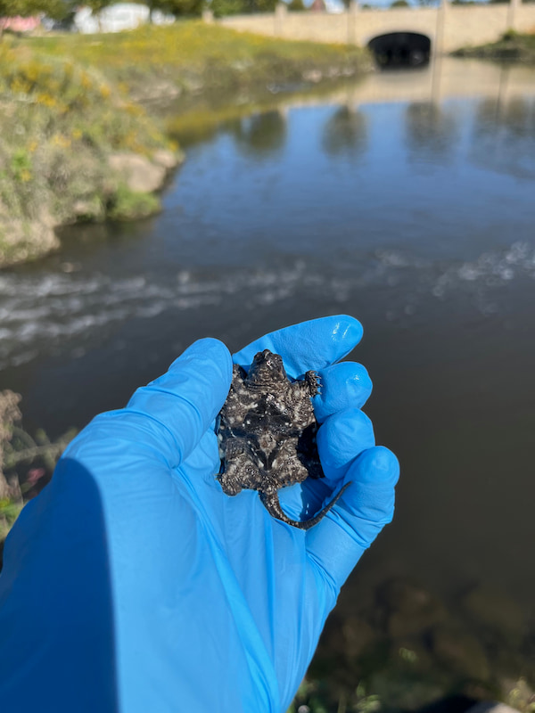
A baby snapping turtle is being handled gently in a gloved hand

If you ever see a turtle on the road, it is very important to help the turtle get to the other side in the direction that it was going. If you decide to pull over, please prioritize your own safety! Not only do turtles have exceptional navigational skills, but they are also very stubborn! Redirecting or relocating a turtle can be very dangerous for them. They may take a longer and potentially more treacherous path to the place they originally wanted to go. Additionally, turtles learn to live and love their home, making it very difficult to learn how to navigate, forage, and socialize in a new environment.

When encountering turtles that may be dangerous to handle, like Common Snapping Turtles, there are still some steps you can take to help! Many wild turtles will be frightened at human interaction and may try to bite. If you are uncomfortable handling any turtle, you can use a car mat or shovel to put under the turtle and slide it. Please never pick up a turtle by their tail, as this can result in severe spinal damage!

What if I find a turtle that’s been hurt?

Turtles are insanely hardy and can survive some pretty horrific injuries. If you come across an injured turtle or one that has been hit by a car, we always recommend bringing it into your nearest wildlife rescue, such as the WMC! If you are unable to bring it in, please contact your local fish and wildlife department to ask for guidance. Never attempt to treat the animal on your own, as this could put you or the turtle in danger and may be in violation of state laws. 

Case example!
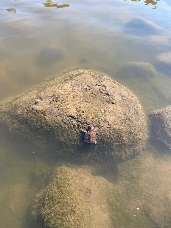
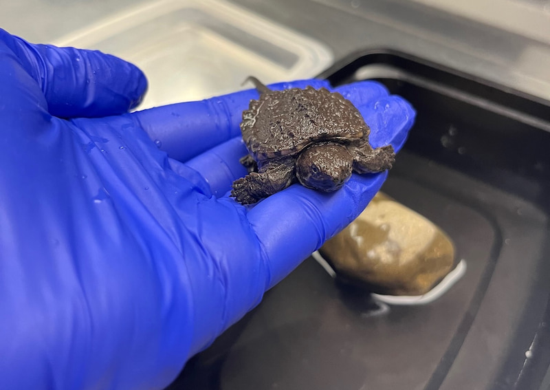
A baby snapping turtle is holding onto a rock outside after release

In mid-September, I was presented with a hatchling common snapping turtle on triage. Generally, snapping turtles lay eggs during May and June, with the hatchlings emerging around September. Therefore, this little one was on perfect schedule! The hatchling had been picked up by a Good Samaritan that had found it trying to cross the road. They were most likely trying to get to the nearest body of water. 

On exam, it was a perfectly healthy baby weighing in at 10 grams! Its age was determined not only by size, but by how squishy the hatchling’s shell was. It also had a visible umbilical scar, as seen in the featured image! In the early stages of life, it is normal for a turtle’s shell to be soft and pliable. This allows for “give” as the turtle expands and grows. Sadly, this is also a major reason for predation in newly hatched turtles. The umbilical scar, or yolk sac, can be thought of as a belly button. It is where the turtle embryo was attached to the shell when it was in the egg. Within about ten days, the yolk sac is fully absorbed and there will no longer be evidence of a scar. 

Time for Release!

In this case, we did not know where the turtle had been found and picked up, so we were very hesitant to release it. We knew that it may be hit by a car or predated upon trying to find its way back to its original destination. It was going to take some deep detective work to find out where the little one had come from.

In the meantime, we set up the hatchling with a proper enclosure in the clinic. This included a tank filled with water, a heat lamp, rocks for a basking area, a UV lamp, and fake plants and logs for hiding spots to decrease stress. We also began experimenting with different food items to see what the turtle would enjoy eating. Some of the options included cut up brine shrimp, turtle pellets, bloodworms, mealworms, and pinky mice. We learned that the patient really took a liking to the brine shrimp!

With a lot of luck and determined investigation, we were able to find out where the patient had been found. It just so happened to be a couple of blocks from a large body of water! A few days after the turtle presented to the clinic, I was able to release it. I hope that it will grow for years to come and be a beneficial contributor to our ecosystem!

Written by: Erika, Class of 2027
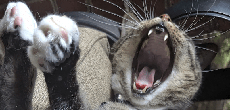
That Bites!

Bite Wounds

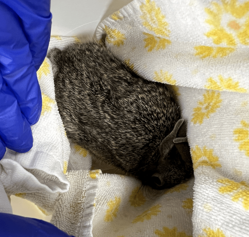
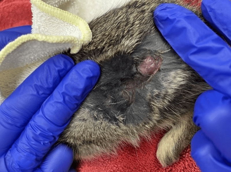
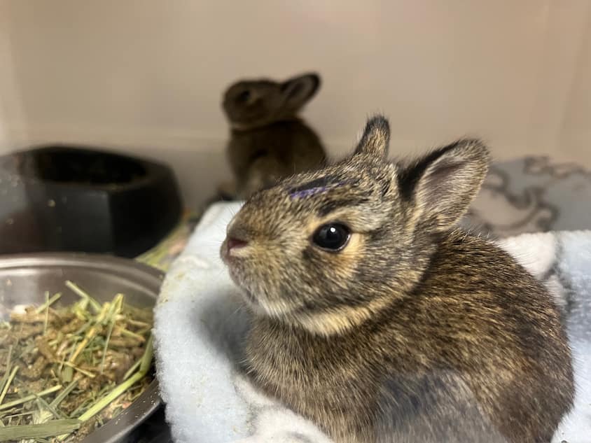
Initial presentation of an Eastern Cottontail with a bite wound from a cat

The Wildlife Medical Clinic has indeed seen it all in terms of injuries from human, domestic animal, and wildlife sources. Whether it be an accident or by instinct, all injuries, big and small, can become lethal to an animal if not adequately treated. One source in particular can be silent but deadly, wounds from a cat’s mouth.

A wound from a cat bite can harbor up to thirteen bacterial species from feline saliva. Most bites can host a mixture of multiple aerobic and anaerobic bacteria that can worsen puncture wounds and prevent healing or lead to sepsis. Several studies discovered that Pasteurella multocida is the most common pathogen within cat bites and is known to accelerate infections. At the Wildlife Medical Clinic, bite wounds are treated immediately with antibiotics and monitored very closely for any sign of infection that can indicate a difference between a healthy release and sepsis.

Case Correlation

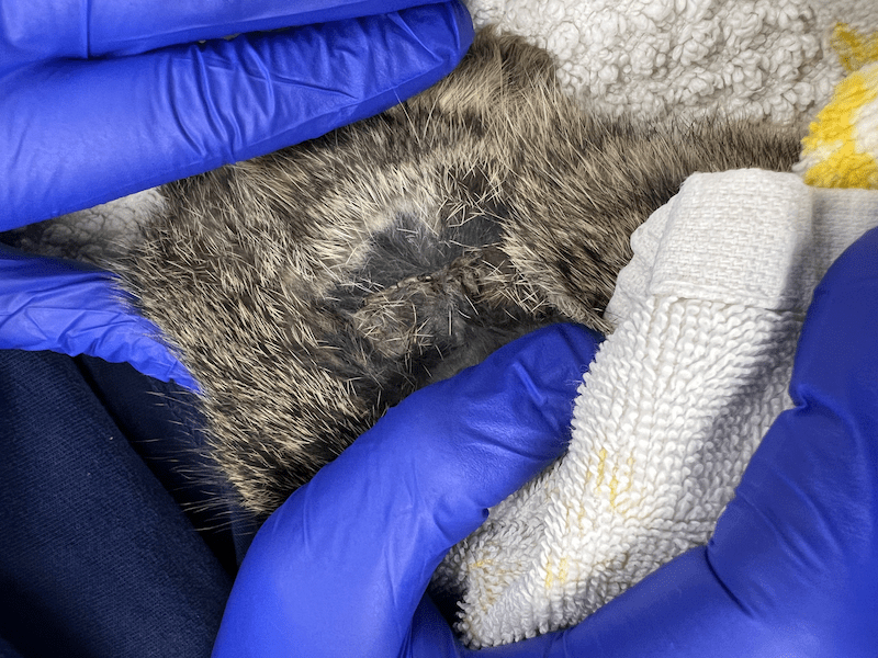
The Wildlife Medical Clinic recently treated a juvenile eastern cottontail after it was bitten by a cat. Upon examination, a large 1.5 inch by .5-inch de-gloving, which is a wound where skin is pulled off the underlying muscle and tissue. Also, a wound was found over the left stifle (knee), but it did not appear to enter the abdominal cavity.

Because of the very thin and friable nature of wild cottontail skin (imagine wet tissue paper), it was decided that any attempt to clip, clean, or suture the wound could cause further damage. Additionally, bite wounds should not be sutured closed, as that can trap dangerous bacteria under the skin. A simple flush of the wound was performed to remove excess debris.

A few other small abrasions were noticed but ultimately were of no concern. The eastern cottontail was ambulating (moving around) appropriately, was of a healthy weight, and had only mild dehydration. We decided that the best course of treatment was to provide fluids, pain medication, and antibiotics. To minimize stress and worsening of the wound, we were hands off as much as we could be with this patient. 

The Outcome

Young rabbits can heal their wounds with impressive speed, and several days later, the wound began to show signs of healing. With no signs of infection or pain, antibiotics and pain medications were discontinued from daily treatment with the plan to continue to monitor. Within eight days, the wound was fully scabbed and posed no threat to normal movement. The eastern cottontail was of healthy weight and was cleared for release! Another successful and rewarding case at the Wildlife Medical Clinic at the University of Illinois!

References:

Talan, D. A., Citron, D. M., Abrahamian, F. M., Moran, G. J., & Goldstein, E. J. C. (1999). Bacteriologic analysis of Infected Dog and Cat bites. New England Journal of Medicine, 340(2), 85–92. https://doi.org/10.1056/nejm199901143400202 

Dire, D. J. (1991). Cat bite wounds: Risk factors for infection. Annals of Emergency Medicine, 20(9), 973–979. https://doi.org/10.1016/s0196-0644(05)82975-0

Written by: Madison, Class of 2027
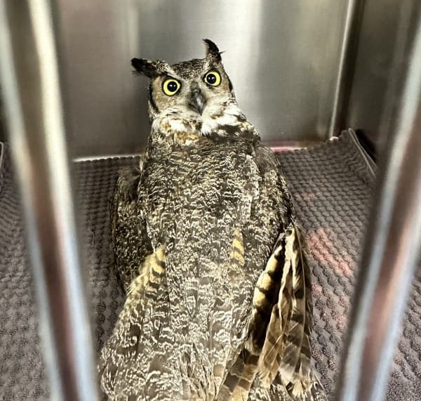
The Rehabilitation of one Lucky, Unlucky Great Horned Owl.

Presentation to the Wildlife Medical Clinic

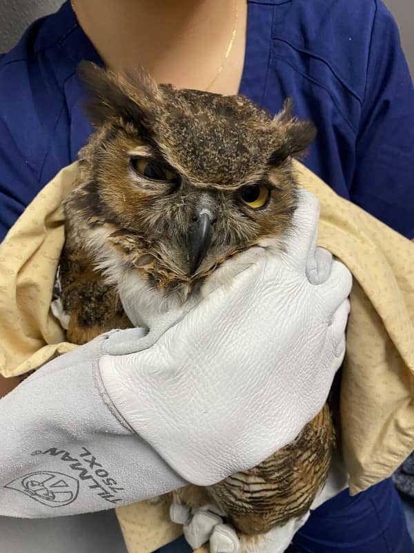
Great Horned Owl in hand upon intake to the Wildlife Medical Clinic.

Bright and early on a Sunday morning, a good Samaritan made an over hour long drive to bring an injured Great Horned Owl, Bubo virginianus, to the WMC. The owl was reportedly found sitting on the highway the night before and was secured in a cardboard box until admission. On triage exam, the owl was found to be standing abnormally on the hock joints instead of perching on both feet but was otherwise bright, alert, and responsive. Upon further examination, it was found the abnormal stance was likely due to severe bruising, swelling, and abrasions to both feet and the left hock. The patient was reactive on examination of the left wing and dark purple bruising was noted at both elbows and the left carpal (wrist) joint. The owl’s eyelids were sunburnt and oddly, all the tail feathers were missing without any sign of trauma to the area. Ocular examination noted hyperpigmentation (darkening) on the ventral aspect of both eyes but no sign of active vitreal hemorrhage (bleeding in the eye) apparent. The owl was also quite well conditioned and was assigned a three out of five score, which is a good body condition score.

After examination, the patient was provided warm fluids, anti-inflammatory and pain relief medications to help with pain, and the injuries over both feet and hock were thoroughly cleaned. Though no fractures were palpated in the wings or feet, examiners wanted to ensure the patient was as comfortable as possible and did not suffer further injury in case a fracture was missed, so the left wing was secured in a figure 8 bandage and the patient’s enclosure was lined with thick fleece until a veterinarian was able to confirm the exam findings and radiographs could be completed. Bloodwork was completed and all findings were within normal limits.

Veterinary Exam, Radiographs, & Ongoing Treatment

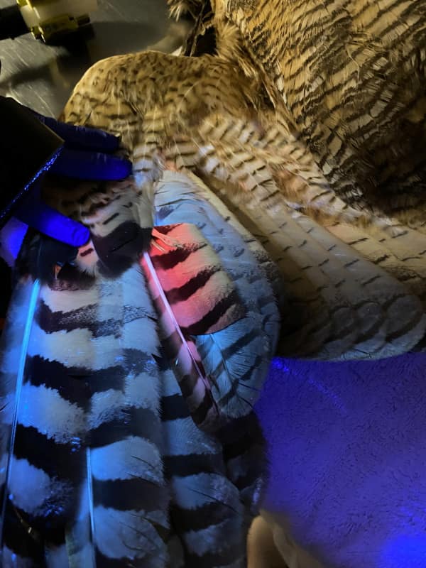
Aging a Great Horned Owl but running a special light over its wing feathers.

Upon veterinary exam, much of the soft tissue damage to the feet had resolved, but bruising to the left carpi and both elbows were still present. Examination and radiographs confirmed there were no orthopedic abnormalities, despite the presumed traumatic event this owl had endured. Subcutaneous emphysema (air under the skin) was noted bilaterally on the flanks (sides) and ventrum (abdomen) but was not severe enough to impair respiratory function. This was one lucky owl! It was decided then to provide supportive care until the soft tissue damage resolved and this patient could be transferred to a long term facility to regrow the rectrices (tail feathers).

During this time, we were also able to age the owl using a Wood’s Lamp. Owl feathers, when they first grow in, contain porphyrins that fluoresce pink under UV light. As the feather is exposed to sunlight and ages, those porphyrins fade, leading to less pink fluorescence under UV light. Owls do not grow new feathers every year, except for their first set of feathers when they are nestlings. Given all feathers did not fluoresce the same hue, it can be inferred that the owl has lived long enough to go through at least one molt of adult feathers, indicating it is at least one year old, a true adult.

For supportive care, the patient continued anti-inflammatory and pain relief medications that were given in a food item during feeding. Treatment teams were instructed to monitor the patient’s mentation, ambulation, and for any injury to the carpal joints. We can also place padded bandages to the carpal area of the wing, called carpal bumpers, to give added protection to that area while the bird is housed in our hospital caging to help avoid further damage to the carpal joints. This was done in addition to fleece padding being added around the enclosure perimeters until the patient was approved to move out into our flight cage.

Reconditioning & Transfer to another Licensed Rehabilitator

When moved out to the flight cages, the patient was placed on the floor to observe flight capability. The owl was able to fly to the nearest perch with significant lift, so it was noted on track for reconditioning.

After ten days in care, this owl was successfully transferred to another licensed rehabilitator to continue flight reconditioning and give it time to grow back all its tail feathers. Prognosis for feather regrowth is fair to guarded given there may be damage to the underlying follicles when they are traumatically plucked, but only time will tell. We are hopeful that this individual will make a full recovery and be released back into the wild.

Written By: Toni, Class of 2027
2024 Year in Review

Happy New Year!

Wildlife Medical Clinic has successfully closed our 2024 chapter and opened our door to 2025. We are grateful for the support from our community, professional colleagues, fellow wildlife rehabilitators, student volunteers, and generous donors. With the collective force from everyone, we were able to provide the best possible care to the injured and orphaned wildlife we received. Without a doubt, our New Year resolution is to continue making a difference by providing excellent medical care for our current and future patients.

We wanted to share with you all the patients we cared for in 2024…

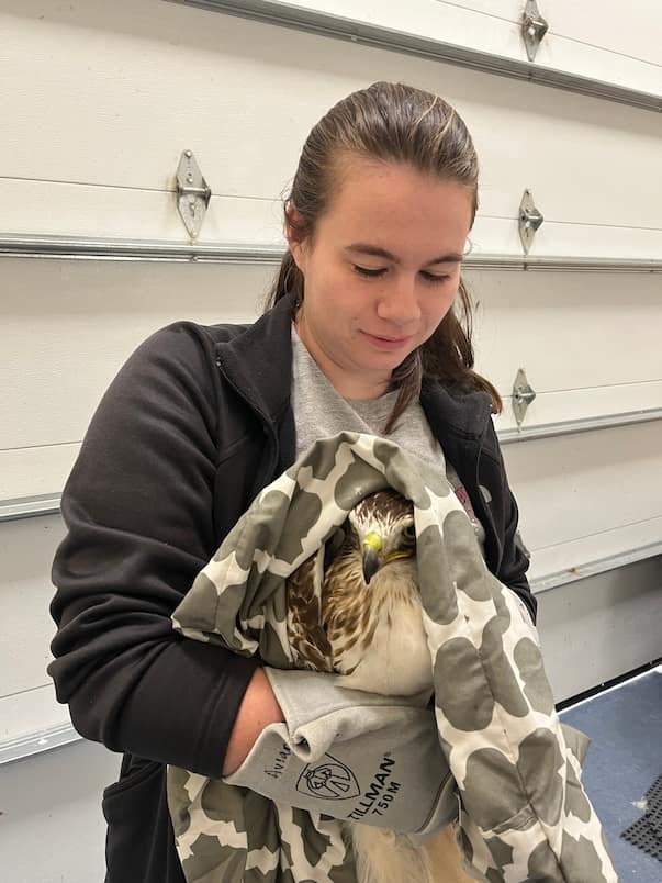
Wildlife Medical Clinic volunteer Alexa, restraining a red tailed hawk.
  • In total, our clinic saw a record-setting 2,278 patients come through our doors, comprising a remarkable 131 different species.
  • Of those 2,278 patients, we treated 7 amphibians, 46 reptiles, 768 birds, and 1,457 mammals.
  • Our most common species in the clinic was…
    • The Eastern Cottontail! They made up 580 of our patients last year.
  • The most common avian species seen…
    • The Canada Goose, which enumerated 93 of our patients in 2024.
  • Our other commonly treated species included the Eastern Grey Squirrel, Virginia Opossum, Raccoon, American Robin, and Red-tailed Hawk.

Thank you all for your continued support of our clinic and the work that our wonderful team does every day to care for all these critters! We are excited about the new opportunities ahead and we will continue to provide care and services to our local wildlife with great passion.

Cheers to continuing to make a difference in the lives of Illinois wildlife in 2025!

Written By: Maddie, Class of 2028

Red-Tailed Hawk Case

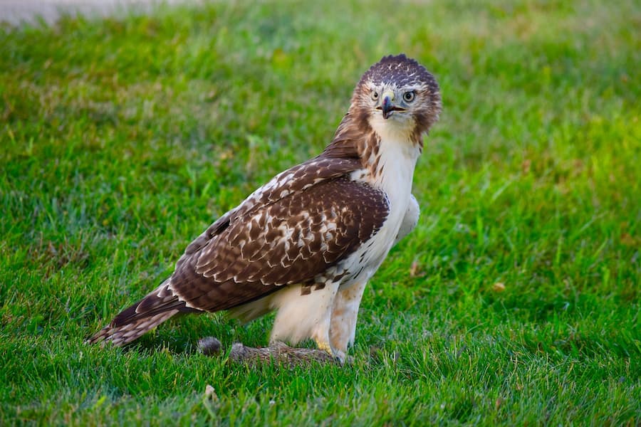
Juvenile red-tailed hawk 24-2042 arrived on September 14 with an ulnar (wing) fracture and ocular (eye) trauma. We provided pain relief medications, topical eye drops, and managed the ulnar fracture with a figure-8 bandage. Once healed, this patient was conditioned for release through a process called creancing.

Nutritional Secondary Hyperparathyroidism

What is Nutritional Secondary Hyperparathyroidism (NHSP)?

Nutritional secondary hyperparathyroidism (NSHP) is unfortunately a common type of metabolic bone disease that can occur in any animal but is most often seen in reptiles and amphibians. This condition most often occurs in captivity when an animal is not receiving the proper husbandry. NSHP can occur when the animal’s diet does not have enough calcium, or vitamin D3, or in the case of many reptiles when they do not receive enough UVB (ultraviolet B) light. Vitamin D3 is required by the body for calcium absorption, so as a result the parathyroid gland, which is found in the neck with the thyroid, begins to produce more parathyroid hormone (PTH). The primary function of the parathyroid gland is to tightly maintain calcium levels and keep them balanced. As a result of the decreased calcium and lack of vitamin D3 the secreted PTH stimulates osteoclasts in the bones, where most calcium is found, to begin resorbing the bones breaking them down causing the calcium to be released back into the bloodstream. Calcium is extremely important throughout the body where it is required for nerve transmission, muscle function, blood clotting and a multitude of other vital bodily functions. While the calcium needs to be in the blood it does so at the expense of the bones.

Why do we see NSHP in Wildlife?

When the bones are broken down to provide calcium it causes them to lose density making them more susceptible to injury. Additionally, there are developmental deficiencies associated with growth that can occur too. All of these make the lives of animals very difficult. NSHP is not as prevalent in the wild, because the animals tend to get enough sun and eat a proper diet. There are two main reasons we see this, first is when people have a pet and they don’t do proper research on housing and diet requirements and therefore don’t meet the needs of their pet. The second reason we see this is due to well-meaning individuals finding wildlife and then proceeding to care for them or keep them in captivity instead of bringing them to a licensed rehabilitator. Wildlife requires specialized diets and housing that are hard to meet in captivity and while people try their best, they often fail to meet the needs of these animals and don’t realize that for most wildlife, permits are needed to care for them, so keeping them in captivity is actually illegal if you don’t possess the proper permits. To further illustrate the effects of NSHP, we will review two cases we have recently seen in the Wildlife Medical Clinic.

Virginia Opossum Case

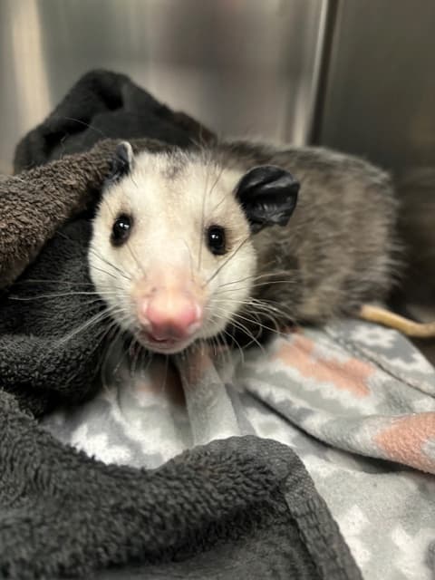
Virginia Opossum with non-symmetrical face due to metabolic bone disease.

On September 9, 2024, we received a juvenile male Virginia opossum that a finder found in their yard struggling to walk. Upon exam he struggled to walk and lacked coordination in his back legs. Additionally, his face was swollen and asymmetrical. Due to the possible orthopedic issue and significance of ataxia we opted to take radiographs.

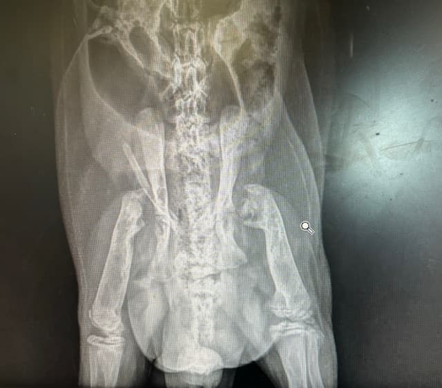
Virginia opossum x-ray.

The radiographs showed that the opossum had a decrease in bone density, and severe malformations of the long bones primarily at the femoral head which is the part of the femur that connects to the hip. They were so degenerated that they appeared to be absent. All the joints appeared to be affected in some way, but that was the most notable location. These changes to the bones and the facial asymmetry led to our diagnosis of nutritional secondary hyperparathyroidism. Our belief is that he was kept as a pet for a significant amount of time when he was younger by someone who did not have the proper knowledge or credentials and then he was released. He also had a very high body condition score indicating he ate well, which would not have been possible on his own, in the wild, due to his musculoskeletal abnormalities. Even though he ate well, he did not receive the proper nutrients needed for bone formation and maintenance. Unfortunately, due to the severity of the malformations and the fact that multiple joints were affected the most compassionate care we could provide was humane euthanasia. He likely had arthritis, which is painful and overall had a poor prognosis for surviving in the wild as the damage done was irreversible.

Common Snapping Turtle Case

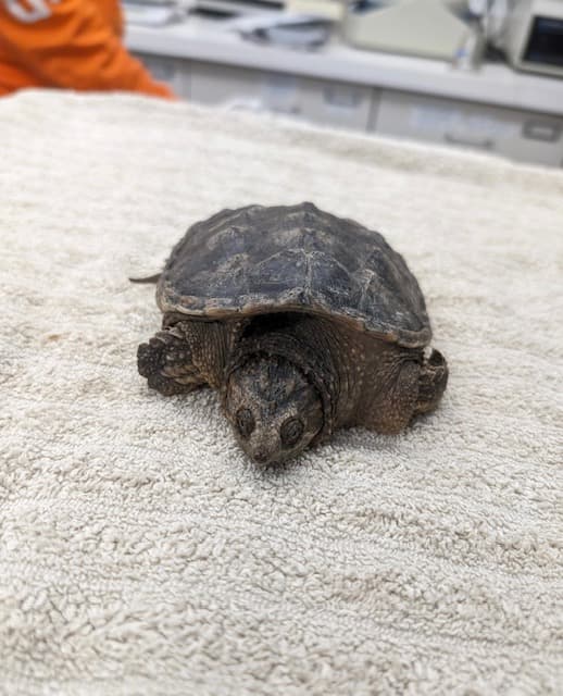
Common snapping turtle on exam table for initial exam after arriving to the Wildlife Medical Clinic.

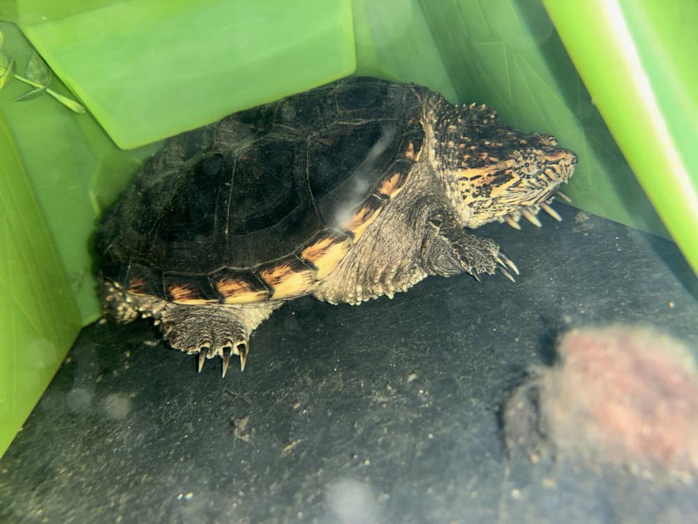
While the Virginia opossum did not have the outcome we hoped for, our next patient, a common snapping turtle, has a more positive outcome. On November 11, 2023, we received a juvenile male common snapping turtle after it was found abandoned in a vacated apartment where he was owned for around two years. He was found dry docked and severely dehydrated. Upon physical exam he was severely dehydrated, his shell appeared too small for his body and was soft and flat. Additionally, he had signs of stunted growth. All of this points to NSHP, primarily the softening of the shell and stunted growth.

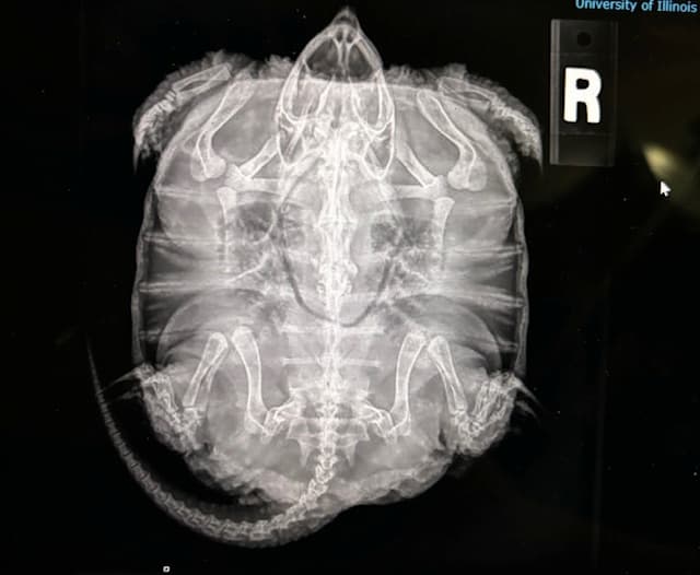
Common Snapping turtle x-ray

This was confirmed through radiographs, which showed decreased bone density and stunted growth Fortunately, unlike the opossum the damage was reversible. He was put on a proper diet along with consistent UVB and heat and now just over a year later he has grown a considerable amount and his body condition has greatly improved. While this is great news, he still is not releasable due to his friendly nature. Because of the time he spent in captivity, he is habituated to people. We have been working on finding him a permanent home and have received interest from an out of state nature center. However, if for some reason this home doesn’t work out, we will keep looking.

Lessons Learned from these Cases

These two cases illustrate the importance of nutrition and proper care for animals. If you want to own a pet, make sure you do the proper research and have everything set up and ready for them before you bring them home. Picking out a pet should not just be done on a whim. They are living creatures that require care and work. The snapping turtle shows the importance of doing research and being committed to owning a pet. Obviously, we don’t know the circumstances of his abandonment, but this is unfortunately an all too common occurrence when it comes to reptiles. Reptiles are unique in their dietary and husbandry needs, which need to be known by the owner, or else serious health issues like metabolic diseases can arise. The case of the Virginia opossum illustrates the importance of knowing who to contact, and only taking care of wildlife if you have the proper knowledge, credentials, and permits. The WMC is built on the idea of us working with the public to better the quality of life for wildlife. If you see an animal that you think needs help, don’t try to keep it and care for it on your own, because it can put you and the animal in danger. Contact a licensed rehabilitator or reach out to the Wildlife Medical Clinic and we can help provide guidance on what to do and who to contact. We hope these cases illustrated how important animal nutrition and husbandry is, why it is best to leave wildlife in the wild, and if help is needed to please contact a licensed rehabilitator.

Written By: Dylan, Class of 2026

Taking Time to Digest

Arrival of Red-Tailed Hawk 24-2154

While the patients presenting to the Wildlife Medical Clinic are typically injured or ill in some shape or form, we do sometimes receive patients that are clinically healthy. This was the case for a juvenile red-tailed hawk who was brought to the clinic on October 3, 2024. This hawk, patient number 24-2154, was brought in after it was found by the side of the road, unable to fly. The finder noted that the hawk still wasn’t moving but seemed to be improving during the drive to the clinic. When we did our initial physical exam on the hawk, everything looked normal, with no signs of any wounds or fractures that might have contributed to its inability to fly. The only thing we found was that the hawk had a very large and full crop. Why would a full crop cause this hawk to not be able to fly? Let’s take a brief look at the anatomy of a bird’s digestive tract to find out why.

Avian Digestive Tract

Birds have a slightly different digestive system than mammals do, especially when it comes to their esophagus and stomach. In general, food will pass from the mouth through the esophagus and its associated crop before entering the two-part stomach and then moving into the intestines. The crop is an out-pouching of the esophagus that is used as a temporary storage location for food. This is an important feature for birds who need to regurgitate their food to feed their young. The storage capacity of the crop is also helpful for birds that want to quickly grab a meal and then move to a safer place to digest it. The stomach of birds is divided into the proventriculus and ventriculus. The first portion, the proventriculus, contains cells that secrete mucus and glands that secrete digestive enzymes and gastric acid. The acidic environment here is especially important for partially digesting bone from the diet, which is an important source of calcium for carnivores. The second portion of the stomach is the ventriculus, which is also known as the gizzard. This portion of the stomach is thicker and more muscular than the proventriculus, and it helps to break down the food into smaller pieces. Many birds will ingest grit, small pieces of sand or stone, to help the proventriculus grind and further break down food material. The ventriculus is also the area where indigestible materials like bones, fur, and feathers are trapped and formed into pellets or casts that will be regurgitated. The ingested material will then pass through the intestines, allowing the absorption of nutrients that the bird needs before it is ultimately expelled as fecal material.

Red-tailed hawk diet

The gastrointestinal anatomy of birds differs between species depending on what type of food they are eating. Red-tailed hawks are a type of diurnal raptor, meaning that they are carnivores who do their hunting during the daytime. In the Midwest, they mostly eat small to medium mammals, such as voles, mice, squirrels, rabbits, and some birds. They are opportunistic hunters, however, and will eat a variety of prey items of different sizes. Red-tailed hawks can swallow small to some medium prey items whole, but in cases of large items, they may instead be taken to a perch to be eaten slowly. These raptors have a crop where they are able to temporarily store food and begin the process of digestion before it reaches the stomach.

Exam Findings

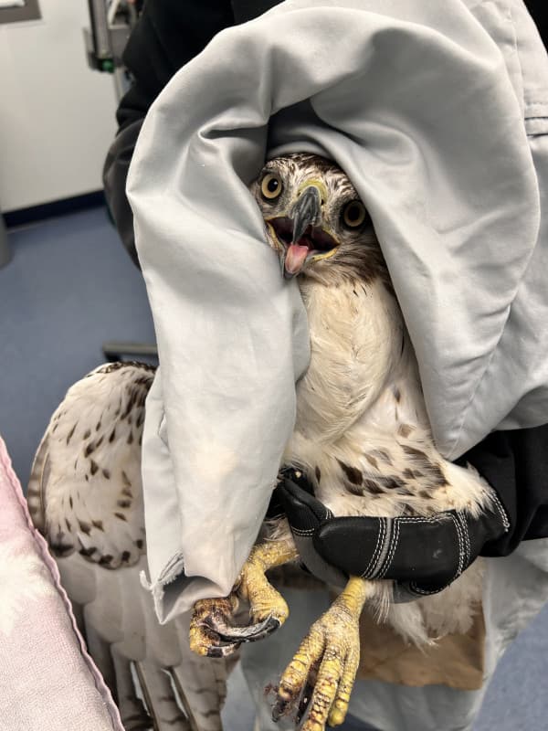
Red Tailed Hawk in hand. Pillow case covering head lifted to see large crop.

Based on its otherwise normal physical exam and large crop size, red-tailed hawk 24-2154 was suspected to have eaten a very large meal that prevented it from flying away properly. The large nature of the crop and the added weight likely caused some balance issues that made it much more difficult for the hawk to be able to take flight. The patient was kept overnight to monitor if it was digesting its prey appropriately and to make sure there were no other signs of injury or infection present. The patient had a DVM exam on October 4, 2024, where the crop size was back to normal, and the patient was completely healthy. The patient was flight tested outside of the clinic on a creance, which is a long weighted line that was attached to equipment placed through removable anklets we placed on the hawk. The patient exhibited normal, symmetrical flight and was approved for release. The hawk was released the following day near where it had been originally found a couple days prior. While this case involved very minimal interventions, this was still a very rewarding case to work on, as the patient made a quick recovery to normal function and was able to be released back to the wild right away.

References:

https://birdsoftheworld.org/bow/species/rethaw/cur/foodhabits

Ritchison, G. (2023). In a class of their own: A detailed examination of avian forms and functions. Springer Cham. https://doi.org/10.1007/978-3-031-14852-1_5#DOI

Written By: Sarah, Class of 2027

Reducing Stress in Wildlife Medicine

Fear Free Approach

In recent years, many veterinary practices have shifted towards a new approach to helping animals feel safe and secure in the clinic. These techniques, called “Fear Free”, aim to decrease stress and fear responses in animals as much as possible, while still being able to conduct adequate exams and treatments. The Fear Free approach aims for a patient-led experience, allowing animals to move about freely with minimal restraint, using relaxing sedatives if needed, and avoiding things that typically induce fear, such as metal exam tables, white coats, and loud noises.

Balancing Act while treating wildlife

In the Wildlife Medical Clinic, creating an environment that minimizes fear and stress is essential for the wellbeing of our patients. In many cases, low stress handling can mean the difference between life and death for wildlife. Uncontrolled stress in animals can suppress immune function and delay healing times, which makes recovery exponentially more challenging for patients. This effect is even more exaggerated in our wildlife friends, as a vast majority of the animals we see in the clinic have no prior experience with humans or handling, and consequently view us as terrifying, dangerous predators. However, this fear response is something that we want to preserve in wildlife, as animals that become too acclimated to humans may not be able to be released back into the wild. Thus, the Wildlife Medical Clinic works to ensure that an adequate balance is upheld when treating wildlife – animals should be handled in a way that creates the least amount of stress as possible, while also ensuring the animals do not become too comfortable around people.

Low Stress Techniques for Wildlife

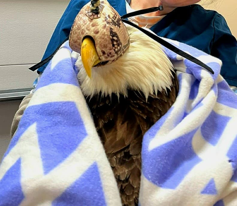
Bald Eagle with hood on for exam.

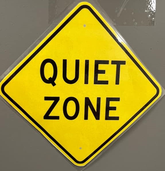
While wildlife patients are in our care, there are numerous strategies that Wildlife Medical Clinic volunteers employ to uphold a low-stress environment for our wild friends. First, direct handling of wildlife species is restricted to only what is medically necessary. This may be for physical exams, medication administration, or wound care. Since this is the most stressful time for our wildlife friends, it’s important to remain hands-off until required. Direct handling of wildlife will always be stressful to some degree, but multiple techniques are implemented to promote a calmer experience while exams are conducted. For example, most bird species, from finches to falcons, calm down when their eyesight is restricted. You will often see volunteers using soft towels or pillowcases to cover the heads of birds, and we even have special hoods for raptor species to minimize stress. Additionally, for extremely stressed patients, sedatives can be administered to decrease the animal’s fight or flight response, allowing for a calmer patient and easier exam. Volunteers also always keep their voices low and calm while handling wildlife, to avoid startling patients with loud noises.

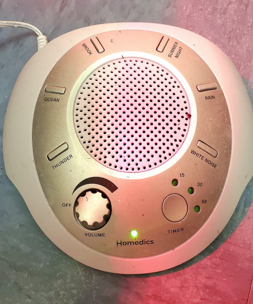
Sound Machine.

You can see low-stress techniques being utilized throughout the Wildlife Medical Clinic, even when animals are not being directly handled. All animals receive their own cages to rest in while they recover. These enclosures can be covered by a towel or blanket to block sightlines and help the animal feel secure and safe in their space. Additional hiding places and enrichment may also be included inside the cages as an added means of security for the patients. Patient holding is located in rooms separate from the rest of the clinic, which helps to minimize the amount of noise and commotion the animals are exposed to. Ambient noise makers are also used inside of these rooms to mimic the natural sounds of the outdoors while they recover.

While being removed from the wild can be quite stressful for our patients, the time and dedication of our volunteers to ensure their stay is as low-stress as possible helps aid in the success of the Wildlife Medical Clinic, allowing animals to recover quicker and be returned to the wild safe and sound.

Written By: Kayla, Class of 2028

Eye-Opening Insights: A Barred Owl Case Update

Barred owl Information

The barred owl (Strix varia) is a larger owl species found in North America. They are part of the family Strigidae, known as the “true owl family.” These brown-gray owls are native to Eastern North America, most often found in mature forests and woodlands. Their main diet is small mammals, but barred owls are known as opportunistic hunters and have been seen consuming other birds, reptiles, amphibians, and even a variety of invertebrates. This is one of the more common owl species that we see here at the Wildlife Medical Clinic.

New Patient Arrival

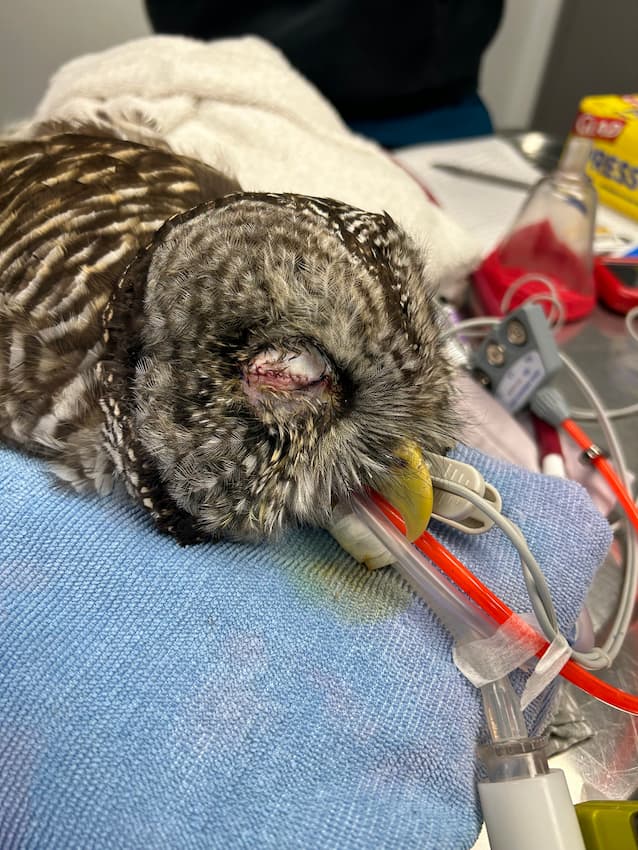
In fact, on August 20, 2024, a barred owl came to the clinic and was in poor body condition, had external parasites, and signs of inflammation and damage in both eyes. During our exam, we found a clavicle fracture that was confirmed with x-rays, which would partly explain why this bird was not in the greatest condition. The clavicle is an integral part of the bone and muscle structure that gives birds the ability to fly. Meaning this individual was not able to hunt for prey due to this injury. These fractures, however, are very easily healed with cage rest. The next issue of concern on the list of problems this barred owl had, was its eye inflammation and damage. We needed to assess whether it had the ability to see. On August 23, we consulted with the Veterinary Teaching Hospital’s Ophthalmology Department, where it was confirmed that in the right eye there was a portion of the retina that was detached, which led to the eye being non-visual. The left eye had some mild inflammation and damage but was determined to be mostly functional. The plan moving forward after this exam was to see if the ongoing inflammation in the right eye would resolve, and if not, the eye would need to be removed via a procedure called an eye evisceration.

Potential Surgical Option for this patient

In general, animals that come to the clinic and are found to have non-functional eyes are given a poor prognosis or ability to survive in the wild. Most of the animals we treat in the clinic either use their eyes for hunting or to watch for predators. Without vision in either one or both eyes these survival instincts are no longer intact, and the animal would either not be able to find food or would be at a greater risk of predation. With owls, due to their nocturnal nature, they rely more on their hearing while hunting at night, than their sight. Because of this, owls are a candidate for the eye evisceration surgical technique to treat non-healing or non-visual eyes.

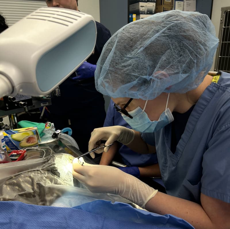
Wildlife Medical Clinic junior student manager, Riley Dunwoody performing surgery on a barred owl.

With this barred owl, the inflammation unfortunately did not resolve, even with medication. Therefore, on the 29th of August, Riley Dunwoody, our junior student manager, performed the eye evisceration surgery under the guidance of our Director, Dr. Lewis. For this procedure, the very front part of the eye, the cornea, is removed and the inside of the eyeball is removed. The outer structure of the eyeball is left intact and the eyelids are sutured closed around it. The outer structure of the eyeball is left intact on purpose. In owls they have a structure known as scleral ossicles within the structure of their outer eye. This gives their face and head the structure it needs to properly allow for sounds to enter the ear. If the entire eye was to be removed, then the face would lose its normal shape, and the owl would lose its sharp ability to hear their prey.

Recovery and Next Steps

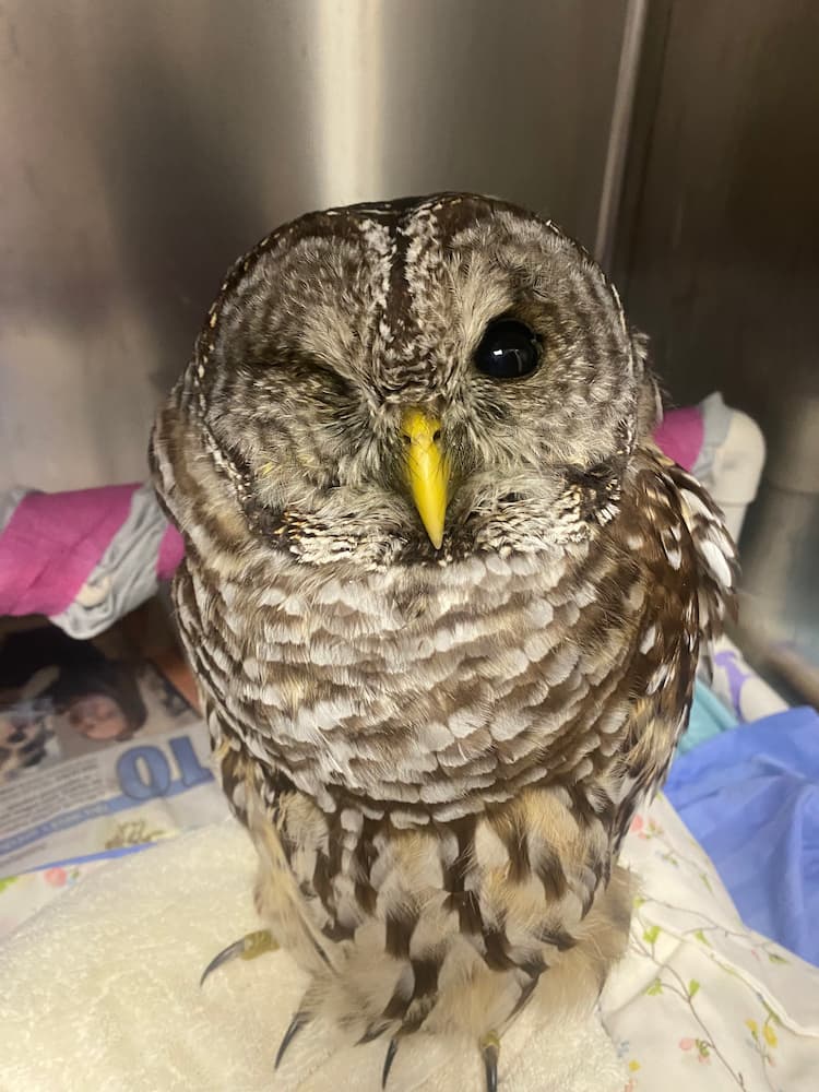
Barred Owl patient in recovery after surgical procedure to remove right eye.

Post surgery, the patient recovered well, their weight significantly improved, and the clavicle fracture showed promising improvement with the formation of a callus (area of extra mineral or bone growth) which indicated the bone was healing. It was decided we would flight test this patient to see if they were ready to be transferred or released. Temporary equipment called removable anklets were placed on the bird’s legs. Then small strings called jesses are placed through the anklets and connected to an extender that is then connected to a creance line. The creance is a light weight, long line that is connected to a weight. This technique can be used to safely evaluate a bird’s flight or to help them build up strength flying to condition them for release. Creancing allows the bird to fly but for a limited distance, usually up to 150-200 feet but we start off at shorter distances and increase the length and number of times they fly on the creance, as they build up strength. However, for this purpose, it was just used to evaluate the Barred Owls flight capabilities. When this bird was creanced, we saw that it had flight capability but would need a lot of conditioning before it was ready to be released. Because of this, we determined that the most beneficial next step would be to transfer to another licensed rehabilitation center that has access to a large flight cage. This would be the best way for this individual to regain strength to fully fly again before it is released back into the wild.

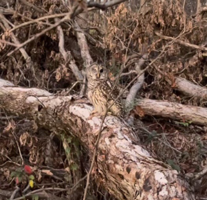
Barred Owl sitting on a fallen log.

After a month with us, on September 24, we successfully transferred this barred owl to a facility with a large flight cage, where it continued its rehabilitation. Our friends at Illinois Raptor Center allowed this patient to recondition in their large flight cages, and on November 19, Riley had the privilege of releasing this patient back in their home territory!

Written By: Riley, Junior Student Manager and Class of 2027

The Journey Back to the Wild

Great Horned Owl, Bubo virginianus

The great horned owl, Bubo virginianus, is found across the majority of North America. Here in Illinois, they are found across the entire state. They are very adaptable and can be found in trees in a variety of habitats, including forests, prairies, and some urban areas. They are easily identified by the tufts of feathers over their ears, which resemble horns.

Arrival to the Wildlife Medical Clinic

Towards the end of August, an adult great horned owl was presented to the clinic by a good Samaritan, and all that was known about it on presentation was that it was unable to fly. On the initial exam, the patient was thin and had severe diarrhea. Radiographs were taken, which confirmed that the patient did not have any fractures. A fecal was run, which showed the patient had a very heavy endoparasite load of capillaria. The patient was treated with a round of dewormers and antibiotics. A creance flight test was also performed, which is where the patient wears falconry equipment and is temporarily attached to a long line that is connected to a weight. Performing creance allows us to evaluate the patient’s flight while being able to easily retrieve them for continued care. At the initial creance, the patient would only defensively posture and did not attempt to fly.

A few days after treatment, a recheck fecal was performed. The capillaria load was lower but still heavy, and another deworming was performed. There is a fungus, Aspergillus, in the environment that birds can be susceptible to if they inhale the spores while they have a compromised immune system. Since there was potential for this patient to have a compromised immune system, a short prophylactic course of an antifungal medication was started as well. The patient had started declining in weight by the end of August and was not as interested in food, so the patient was supplementally fed for several days to prevent any further weight loss. 

Continued Care

A recheck was performed on a fecal sample in early September to re-evaluate the patient’s parasite load, and there were now fewer capillaria seen, but another endoparasite (coccidia) was also seen. Yet again, another dewormer was given for the intestinal parasites. A blood sample was also taken at this time and sent to the Veterinary Diagnostic Lab at the University of Illinois, and it was found the patient had moderate levels of three different blood parasites, including Plasmodium, or avian malaria. At this point, a daily anti-malarial drug called pyrimethamine was started. The goal for treatment for these blood parasites is to get them to low levels, as the patient will never be completely cured of them. A fecal and blood sample were rechecked about a week afterwards to monitor the patient’s response. The initial two intestinal parasites were not present, but another type of intestinal parasite (strongyle) was seen. Another dose of a dewormer medication was given. There were no major changes seen on the blood sample, but the blood parasites that were present can require a lengthy period of treatment to get them adequately controlled. At this time, the amount of food the patient was being offered was also increased to help keep weight on the patient, as all the food offered was being eaten.

Turning the Corner and Gaining Strength for Release

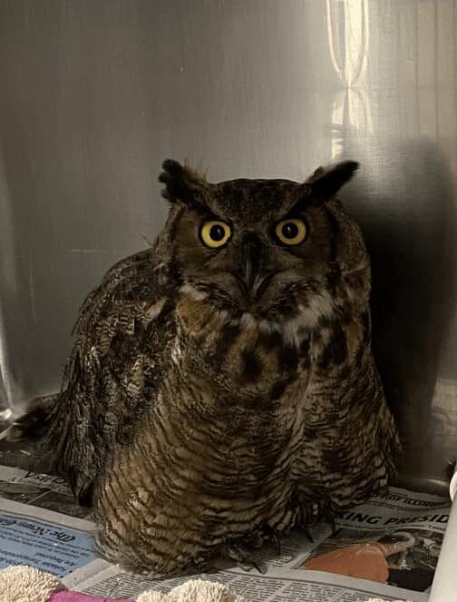
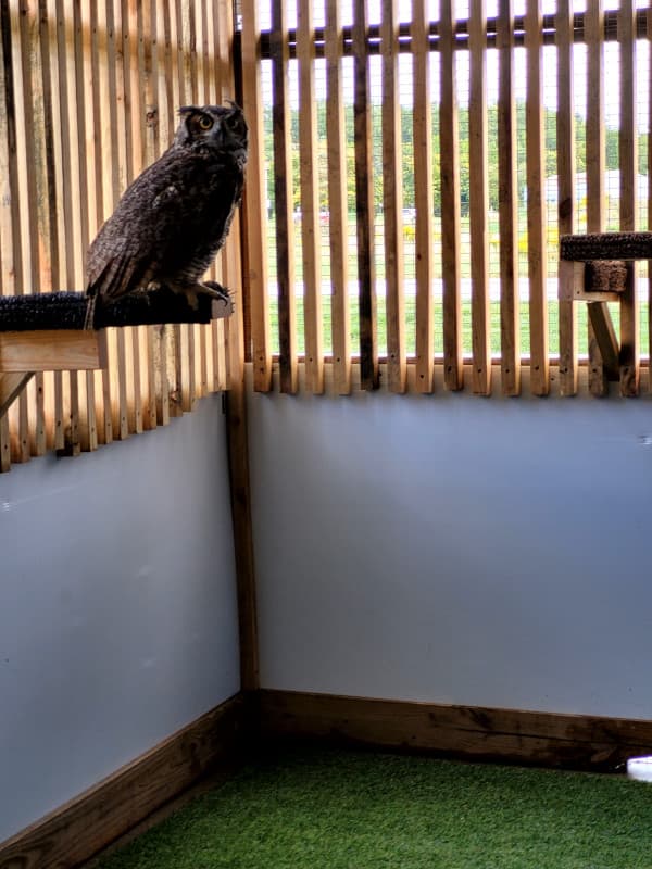
Great Horned Owl patient in our outdoor flight cage. This bird has gone through treatment and is gaining strength back before being released.

Towards the end of September, a recheck was performed where blood and fecal samples were taken and another creance session was completed. The fecal showed a very low strongyle load. It was elected not to administer another dewormer at this time as it was a low number of parasites, and the patient will naturally come across these parasites in the wild. The blood sample showed lower levels of the blood parasites, and it was decided to stop the anti-malarial course the patient had been on, as the levels were low enough for the immune system to keep them in check.

We tried another creance session and this time, the patient flew short distances and had symmetrical flight. This patient was getting stronger and healthier, so it was time to move them to an outdoor flight cage. This allows the patient to move around in a larger space and for us to access their flight better before considering release. After monitoring the patient in the outdoor flight cage, we decided to transfer them to another licensed rehabber in early October to continue monitoring the progression of healing while allowing the patient to fly in an even larger enclosure and gain more strength before returning to the wild.

Written By: Angela, Class of 2026

Wildlife Medical Clinic
John A. Coyne South Clinic
2100 South Goodwin Avenue
Urbana, IL 61801
217-244-1195