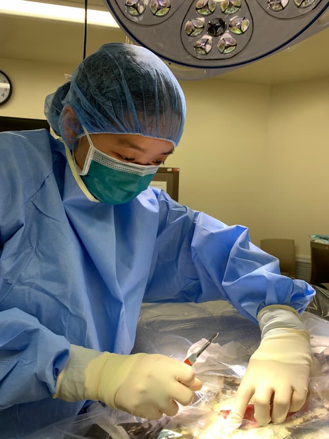
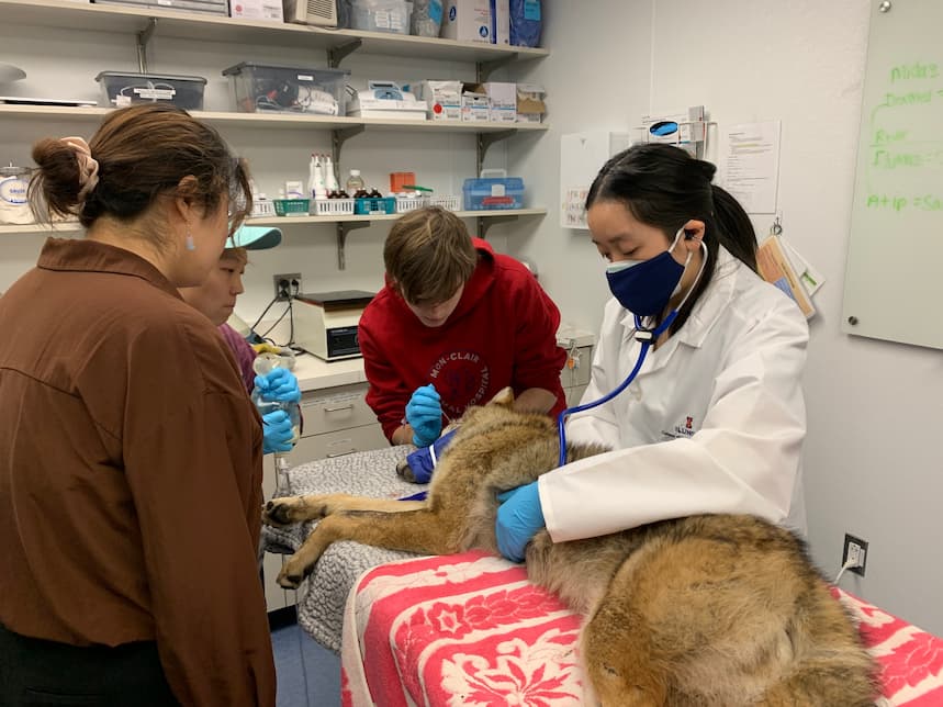
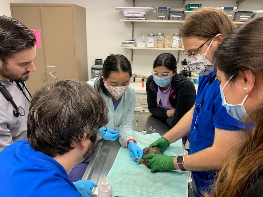
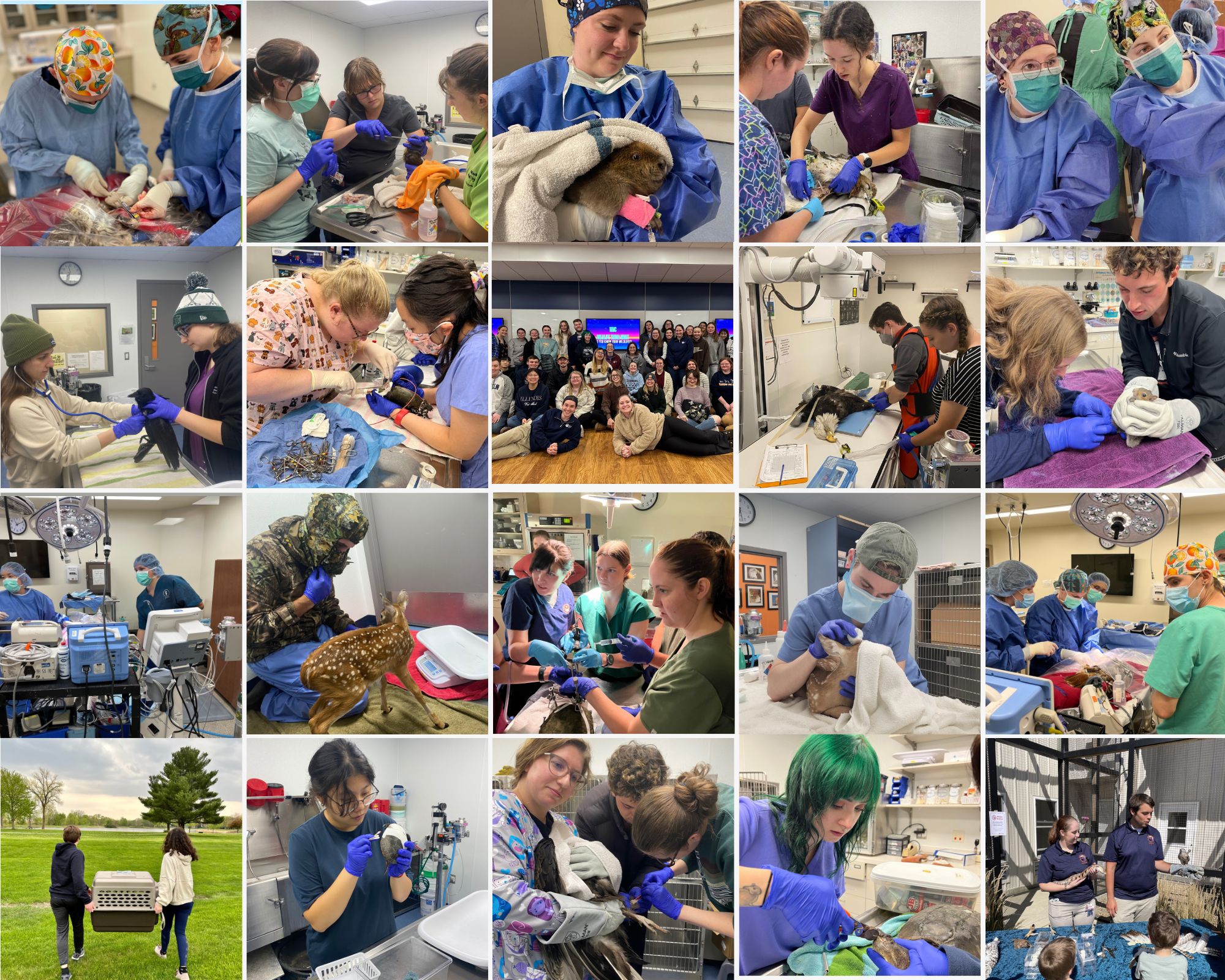
As the summer draws to a close, we are overjoyed to welcome our talented students back to the clinic. The clinic has been working tirelessly to update and revamp our volunteer offerings by bringing in community members, expanding opportunities for students outside of the veterinary college, and improving the learning curriculum for veterinary students.
Students Take the Lead
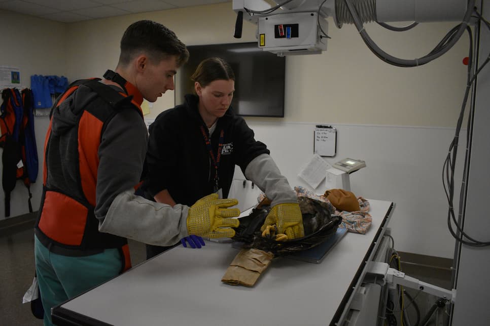
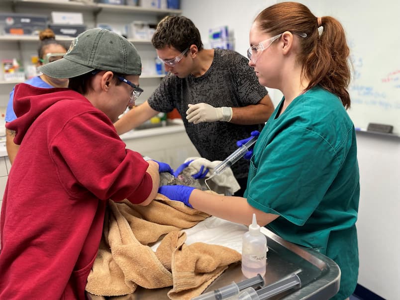
Over the summer, the WMC operates much like a standard wildlife rehabilitation center with a small, dedicated staff and rotating clinical year students. As we transition into the academic year, we return to our unique management style: students take the lead. Students are not just participants, they are integral to every aspect of the clinic, from novice volunteers to upper management. This approach allows students to maximize their learning opportunities and acquire a diverse set of skills beyond wildlife medicine. Even with students at the helm, there is a robust safety net and support system of talented veterinarians, guiding and assisting students every step of the way.
Team Leader Training
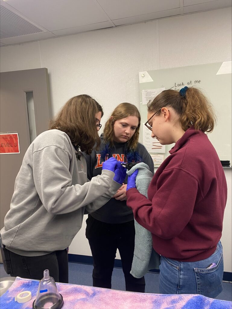
As we kick off the semester, our student team leaders have been diligently training our new volunteers, ensuring they are prepared to handle their own patients and care for our diverse wildlife species. The clinic’s current functioning would not be possible without these dedicated team leaders. Their selfless commitment to improving the lives of the wildlife in our care and the learning experience of incoming students is truly commendable. During their new full-day training, team leaders worked together to set goals for themselves, their teams, and the clinic. They also defined what it means to be a team leader: support, compassion, education, teamwork, and growth. By embodying these five traits, team leaders continue to enrich the lives of WMC students and maintain the highest welfare standards for our wildlife patients.
New Orphan Feeding Team
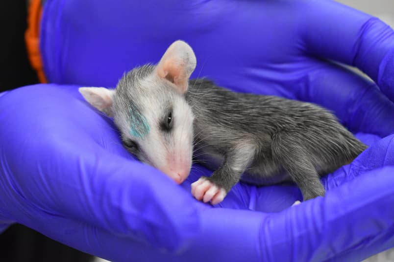
The advent of our new orphan feeding team opens new opportunities for community members and outside students to gain experience in wildlife rehabilitation and orphan care while bringing in more hands to assist with our busy caseload. Orphan care, while highly rewarding, can be time-consuming and overwhelming if there are not enough trained volunteers to help. Our caseload has increased year after year as word of our services has spread, creating a need for more volunteers. We aim to establish the orphan feeding team as a new local volunteering staple and bring the community to us to foster a love of wildlife.
Dedication to Wildlife
As we welcome the new academic year, we are thrilled to have our dedicated students back at the clinic, assuming crucial leadership roles and contributing to the welfare of our diverse wildlife species. The commitment of our team leaders and the establishment of the orphan feeding team underscore our unwavering dedication to providing unparalleled learning opportunities for students and maintaining the highest standards of care for our wildlife patients. We are excited to continue fostering a love of wildlife in our community and look forward to the positive impact our clinic will have in the coming year, as we remain steadfast in our commitment to wildlife care and community engagement.
Written By: Tyson, Senior Manager and Class of 2026
Summer is the busiest season in the Wildlife Medical Clinic! To ensure we provide quality care and support to our patients, our summer interns have been putting their whole heart into caring for our wildlife patients and supporting our faculty, house officers, and 4th-year vet students. Each year, we hire 3-4 veterinary medicine students, who have been involved in the clinic for at least a year, as summer interns. Their main responsibilities are to provide near-peer mentoring to our 4th-year students on case management, medical treatment, diagnostic choice, and all things wildlife. Below are some of the case highlights from each of our summer interns this year and their reflections on the season!
Rachel Eggleston, Class of 2027- Case Highlight:
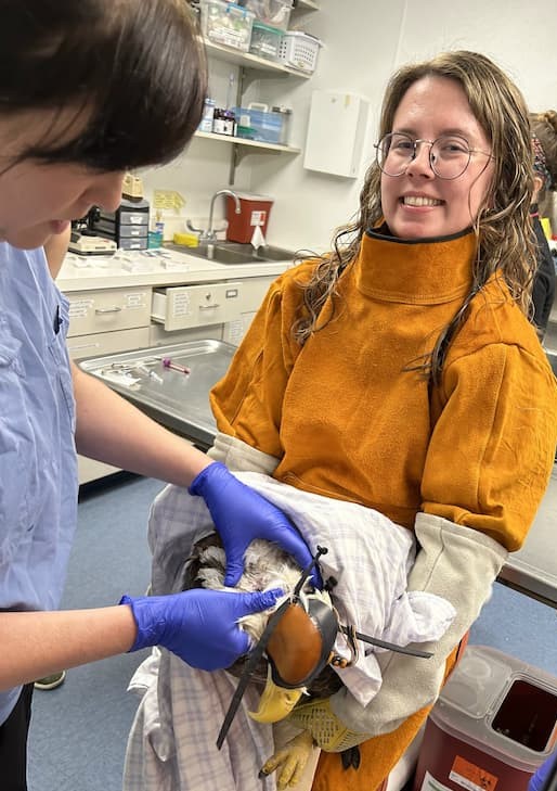
At the beginning of August, the Wildlife Medical Clinic received a female American Kestrel. She came to us after being found near the side of the road and was not acting normally. I do not have a lot of experience with American Kestrels in the clinic and I really enjoyed getting to be involved during the triage exam for this patient. During her physical exam, there were not any obvious injuries or abnormalities, but we did notice some increased laxity in her right wing. We also looked into her eyes with an ophthalmoscope and were able to see evidence of minor hemorrhage or blood in the right eye. While she has been in the clinic, we have been providing pain medications, anti-inflammatories, and additional supportive care as needed. I look forward to working with her more and seeing her progression as she continues to heal!
Rachel’s Thoughts
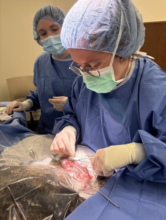
My name is Rachel Eggleston, and I was chosen to be one of the DVM student interns for this summer! I will be going into my second year of veterinary school this fall. Some of my favorite memories from this internship have been getting to work with different raptor species. One of my favorite memories that I will remember for a long time is getting to scrub in and suture close on a surgery for a Great Horned Owl! I have always been a fan of birds and thoroughly enjoyed helping treat a Bald Eagle we had in the clinic for a while before it was transferred to a different wildlife center. I love so many things about being able to work in the WMC, but I think what I value the most from my time this summer is the long-lasting connections I have been able to make. I have greatly enjoyed getting to work alongside clinical year students, faculty, residents, and the WMC managers. This summer program has allowed me to grow and strengthen my skills as a future veterinarian, student, and colleague, and I am so grateful to have had this opportunity!
Roxanne Lisowsky, Class of 2027- Case Highlight:
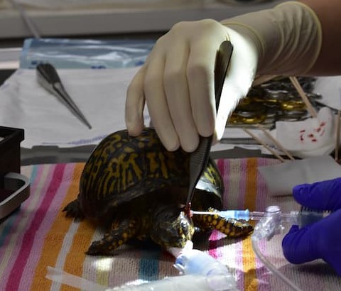
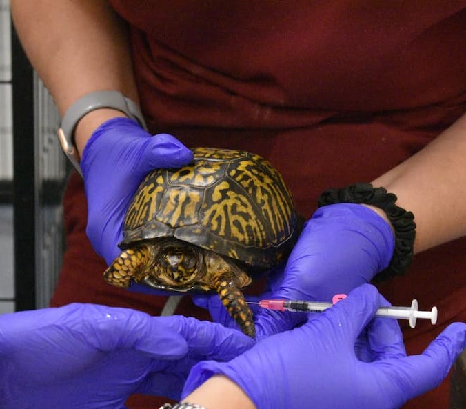
In early July, we had an adult female eastern box turtle come into the clinic for swelling on the sides of her face and not moving around much. Upon our initial exam we found that the patient had aural (ear) abscesses on both sides. The turtle also had some crusting and discharge in both eyes along with nasal discharge. We were able to drain the larger abscesses and started her on antibiotics to help clear the rest of both abscesses. We also gave pain medication and anti-inflammatory medication to help with her healing. During her stay we also found she had corneal ulcers that we treated with eye medication, and they resolved. We had to drain the abscess one more time during her recovery. We also tested for mycoplasma, and it came back negative while the culture of the abscesses came back sensitive to the antibiotic we used. She continued healing and recovering well in the clinic. Turtles heal very slowly, and it takes a lot of patience and dedication to help them along the way. We made sure she finished her course of antibiotics and then she was given one last exam to confirm her clean bill of health before being released on August 9. We hope she has a long and healthy life!
Roxanne’s Thoughts
My favorite part about this summer has been working with the undergrads and 4th-year students. I love being able to show them new things about wildlife patients, teaching new skills, and seeing the lightbulb moments. I grew more confident in my daily clinical abilities and feel prepared and excited to be a team leader for the next school year. I also enjoyed seeing patients we don’t always get during the school year.
Caroline McCormick, Class of 2027- Case Highlight:
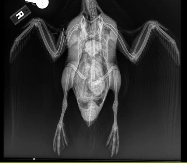
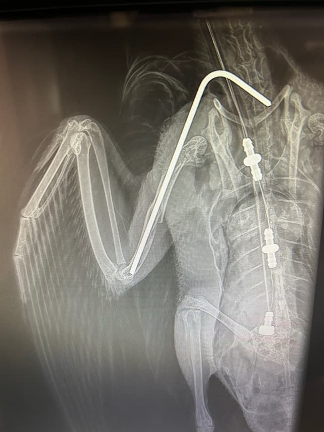
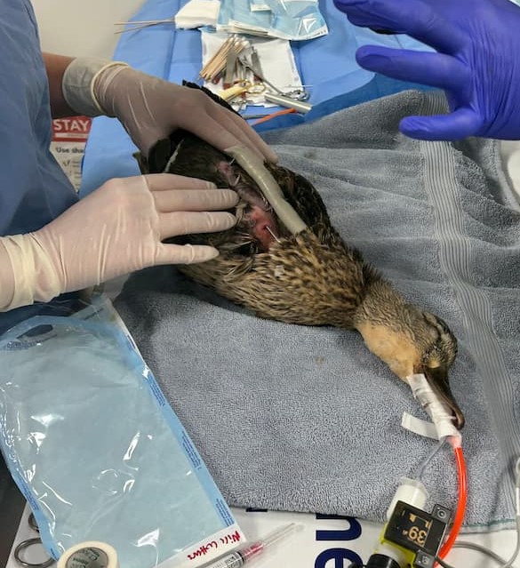
An adult female mallard presented to the wildlife medical clinic on May 29, 2024, with an acute mid-diaphyseal long obliqued simple right humeral fracture, opened on ventral aspect with ~1-2 cm of exposed bone with each fragment. The injury appeared very acute (< 6 hours old), and not grossly contaminated. There was relatively minimal muscle/soft tissue damage.
Immediately upon intake, radiographs were performed to confirm the injury, and the animal was prepped and taken to surgery. An inter-medullary pin was retrograded into the right proximal humerus via existing the ventral wound. The distal fragment was aligned and the IM pin advanced until it was well seated in the distal fragment. The open fracture was closed with a continuous horizontal mattress suture. The IM pin was bent using a chuck and vice grip and then blunted. A body wrap was placed to stabilize the wing for healing. The mallard was then started on pain medications and antibiotics.
Physical therapy was performed three times per week for several weeks to prevent loss of range of motion in the wing. The pin was slowly destabilized or removed as the wound healed. Repeat radiographs were performed to monitor healing. Eventually the pin was removed completely. Once the mallard was healed, she was transferred to another facility for continued rehabilitation before release.
Caroline’s Thoughts
The summer program has been wonderful. I have enjoyed working with so many wonderful people and animals. I have learned a lot and been able to practice many skills.
Sofia Murillo Logan, Class of 2026- Case Highlight:
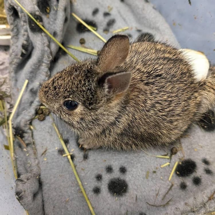
One of the most inspiring cases from this summer is from a small bunny that came into the Clinic. An unidentified juvenile eastern cottontail presented to the Wildlife Medical Clinic on June 14, with a history of a broken left leg. On the intake exam we found a fracture of the right tibia, but other than that, this bunny was pretty healthy. Now when animals come into the clinic there are a list of factors we always need to consider for the health and safety of the animals and the volunteers. “How will this animal respond to captivity while in the clinic, and what is this animal’s prognosis.” Prognosis meaning based on everything going on with the animal and the road they face ahead, how likely are they to survive to then be released.
When speaking to Dr. Lewis about this case and evaluating the prognosis of this patient we decided to keep this patient for further evaluation. Due to their good body condition and the stability of the fracture we created a plan for this patient. Body condition can be a huge aspect to the prognosis of a patient. This comes down to, how stable is this animal, and if it is not stable, how long will it need to become stable. Without the patient being in good body condition and having appropriate neurological signs, it can be hard to treat severe injuries successfully.
Throughout the patient’s stay the bunny was doing very well. We stabilized the fracture site with an Ehmer sling, the same process as when you need to put your arm in a sling when you shouldn’t use it, for healing purposes. We kept the bunny on some pain medications to keep them comfortable while in our care. We took radiographs (x-rays) on the patient to visualize the fractured leg. After confirming the stability of the fracture, the doctors were able to fix the leg surgically by placing an intramedullary pin. This is a pin just a little smaller than the width of a bone that is used to keep the bones in place while the patient is healing. After surgery our patient was still doing well and gaining weight, which again eating is a positive prognostic indicator. After about a week we checked to see the healing progress of the patient by taking another radiograph. Since there was evidence of a bony callus starting to form, we removed the sling and allowed the patient to freely move around the cage to encourage movement of the stifle (knee) and hock (ankle). After another week another radiograph was taken to see if it was time to take the pin out of the patient’s leg. Due to the stability and healing of the leg we did decide to take the pin out. The pin was removed a little over 2 weeks after being placed. We were able to remove this pin this early because bunnies can heal very quickly, especially young ones. We also didn’t want the pin to limit this growing bunny’s leg, so we were very happy to remove it so quickly. The bunny was able to be released a few days after pin removal.
Overall, the patient did very well and gained over 100g during their time with us in the clinic. Something we could have done differently in this case was to keep this patient a little bit longer to get more strength built in the injured leg but ultimately it was doing well, and we felt comfortable with releasing when we did. We were very happy with the result of this case and hope this eastern cottontail is out in the wild eating all the greens and growing to adulthood!
Sofia’s Thoughts
My name is Sofia Murillo Logan and I am one of the Wildlife Medical Clinic Summer Interns. I applied to this internship because I wanted to spend more of my time immersed in wildlife. Volunteering for the clinic in the school year can be difficult due to the rigors of veterinary school. My experience thus far has been nothing short of inspiring. I get to learn daily from our amazing WMC Director, Dr. Stephany Lewis, as well as our house officers that are working towards specializations. Being at the clinic has increased my confidence and knowledge not just in wildlife species, but also in medicine and clinical reasoning. I have greatly enjoyed working with our fantastic and knowledgeable managers Tyson Jenkins and Riley Dunwoody. They both are incredible leaders and are passionate about doing what’s best for all the creatures in our care, as well as for the students in the clinic. For any students at the University of Illinois interested in gaining more hands-on experience I highly recommend getting involved in the Wildlife Medical Clinic.
Written By: YiYing and Sofia, class of 2026, and Rachel, Roxanne, and Caroline, Class of 2027.
Dr. Amanda Wong our wonderful Zoological Companion Animal Resident recently completed her residency at the University of Illinois Urbana-Champaign. She spent countless hours at the University of Illinois Zoological Service and the Wildlife Medical Clinic for the last three years. And many of our volunteer students had the honor of working with and learning from her. She always put her students first and motivated them to take on the challenge of practicing new skills. This July, Dr. Wong left the University and moved on to the next chapter of her career. Although we were sad to see her go, we wish her the best in the next chapter of her career. However, before she left we conducted a mini-interview with her and are excited to share!
Dr. Wong graduated with a Doctor of Veterinary Medicine degree with a background in small animal medicine with an interest in internal medicine and emergency care. Over time, she developed a career interest in zoological companion animals (ZCA), pets that are not small or farm animals. She especially enjoys human and pet ownership in the ZCA world and the potential of medicine, which has a lot of room to explore.
Favorite Memories
She highlighted her interaction with our students when asked about her favorite memories at the Wildlife Medical Clinic. “… working with students, especially during the summer months, were some of my best memories.” Dr. Wong said she was impressed with the student’s potential and ability. “I remember working with Becky (DVM candidate, class of 2026) on her turtle patient. She took the initiative to learn to place the feeding tube. And she successfully performed the procedure on the patient by herself with only verbal instruction!” Another case that left a deep impression for Dr. Wong was a coyote patient that she worked with student volunteers, “… we don’t get adult coyote often. It was a unique opportunity for students and us (house officers) to think about patient and student safety.”
Virginia Opossums are the best!
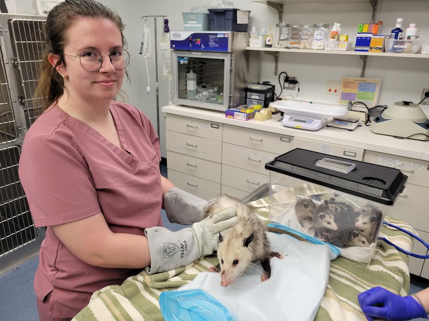
Dr. Wong has worked with hundreds of wildlife patients at the WMC, and opossum is her top wildlife patient to work with. They not only are incredibly charismatic (think baby opossum here), but they are also great for the environment with their fantastic pest control ability.
Future Career Plans
After leaving the University, Dr. Wong plans to return to California’s Bay area. She will be joining a private practice focusing on the zoo companion animal. Her long-term goal is to become a board-certified specialist working in private practice and emergency care and one day open her own practice!
Words of Wisdom
Before we close the interview, she would like to share a word of encouragement with all the readers and our current and prospective students. “WMC, in general, is a fantastic opportunity; learning from the WMC and the cases will serve you very well. I Highly encourage getting involved in the clinic.”
THANK YOU, Dr. Wong, for all of your hard work and the guidance you provided to our students; we will miss you so much! We are sending all the best wishes for your future endeavors.
Written By: YiYing, co-chair and class or 2026 and Rachel, co-chair and class of 2027.
Have you ever been walking along on a sunny day in the spring, going to visit your family, perhaps, when suddenly, you find yourself ankle deep in cement? Or quicksand? Maybe molasses? If you answered “yes” to any of these scenarios, then you will be able to empathize very well with what animals experience when they encounter a glue trap.
The Reality of Glue Traps
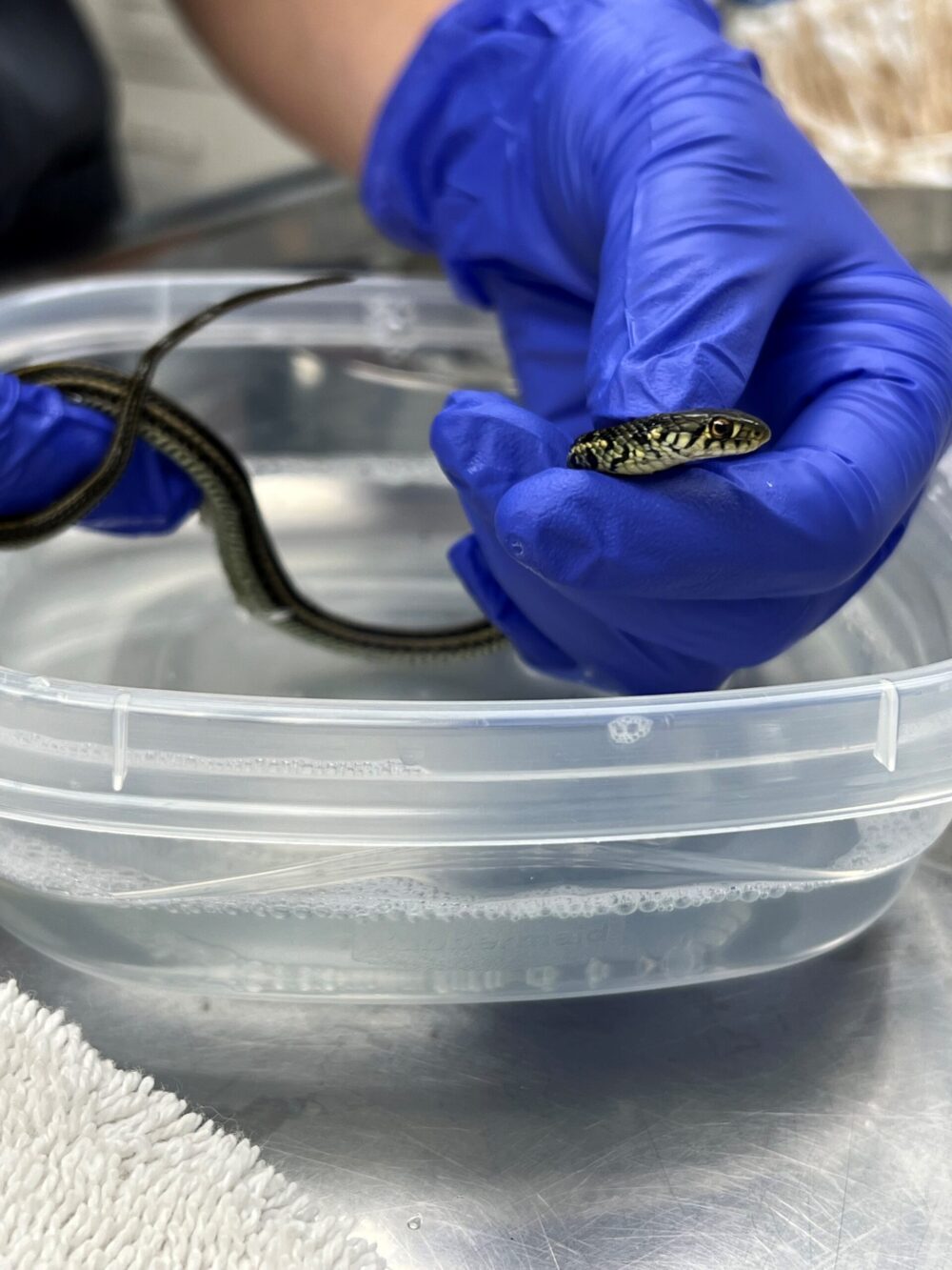
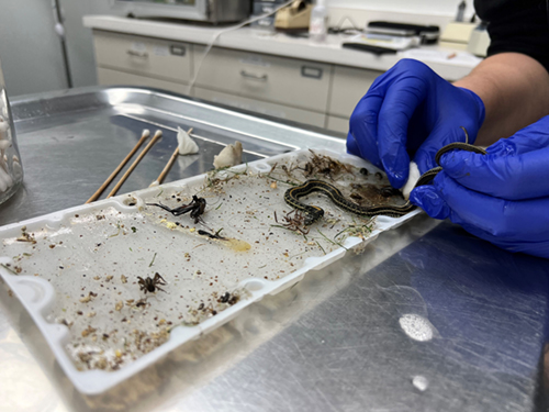
Glue traps are used to prevent animals such as rodents from entering unwanted areas. That is how they are advertised, at least. The reality behind glue traps is far from that simple, however. Glue traps are one of the most inhumane methods of pest control that currently exists, despite being commonly used. They frequently catch more than just rodents; any type of animal can get stuck in one, such as birds, reptiles, amphibians, invertebrates, and even cats if they’re small enough. Just this year, the Wildlife Medical Clinic received several animals stuck to glue traps. Most recently, it was two Common Garter snakes, and they were stuck on the same trap. Thankfully these snakes were brought to us before they experienced any significant state of decline and were eventually released, but many animals are not so lucky.
Most animals that are caught by these traps do not have a quick painless death. They can be trapped for several hours to several days before succumbing to dehydration, starvation, exhaustion, and exposure to the elements. If they are lucky enough to be rescued, many still have lengthy stays at rehabilitation centers due to injuries from trying to free themselves and may need time for feathers and fur to grow back and damaged scales to heal. Thankfully though, there are several effective and humane alternatives!
How we can Help
Here, we can see how wildlife creatures get completely stuck to glue traps.
The best way to keep unwanted wildlife guests out of your home is to ensure that it’s locked up tight and is very uninviting (think, opposite of Goldilocks and the Three Bears). That means storing food in air tight, sealed containers, as well as making sure any cracks or holes leading in are sealed up, especially during the change of seasons. This applies beyond just mice and rats as well; this will ensure you do not have any LARGER guests, such as squirrels or even raccoons, finding your house just a little too inviting.
If you do ever have some unwanted visitors in your home, though, there are solutions to help them find their way out that avoid things like glue traps, or even having to use lethal force most of the time. Different scents, bright lights, and loud music are a few of the options. Basically, make your house unhospitable to your unwanted guests. Strong scents like peppermint will deter them from an area, so applying a few drops on some cotton balls and leaving them at potential entry points around your home (and out of reach of pets and small children!) is a great alternative. Snap traps are another way that you can prevent your home from becoming the new hotspot on the block for wildlife. Other methods such as rodenticides and moth balls are not recommended; rodenticides cause devastating damage to wildlife populations through the food web. Rodents ingest the toxin directly, then carnivores such as raptors, coyotes, foxes, and more, ingest the poison indirectly after they consume what they think is an easy meal, aka, the carcass of a rodent who ate rodenticide. Moth balls are carcinogenic and should not be left out for anyone to be exposed to, humans included!
So, this spring, in the spirit of new life and warmer weather, opt out of the old methods of keeping furry friends out of your house. This is one of the single instances in life where you are ENCOURAGED to be a bad neighbor (play loud music, keep bright lights on, have noxious smells around your home) in order to keep yourself, your family, and also wildlife, safe and free, especially from glue. Happy Spring!
This article was written by third year student Macy (class of 2025)
I have wanted to volunteer with the Wildlife Medical Clinic (WMC) since being accepted into the University of Illinois College of Veterinary Medicine. My goal is to become a zoo veterinarian, and volunteering in the WMC is the perfect opportunity to work with some of the species I might see throughout my career working with exotic animals, or at minimum I will see scenarios and cases that can be applied to others in which I will be involved in the future. I signed up right away with a lot of aspirations for what I could learn from the other students, faculty, and residents at the clinic. I started out semi-confident in my clinical skills as I had some small animal medical experience. I also have a zoology background with husbandry (daily care and needs) knowledge of the animals we might potentially see in the clinic. I was excited to finally combine the two experiences by volunteering in the WMC!
As I worked with the team and learned the ropes, I was able to work on two interesting cases before winter break that taught me a lot about the medical process of rehabilitation and what goes into a successful release. I was the secondary on those cases or the number two in who was making decisions, and I was very grateful that the primary on both cases was such a great mentor as I learned a lot from them.
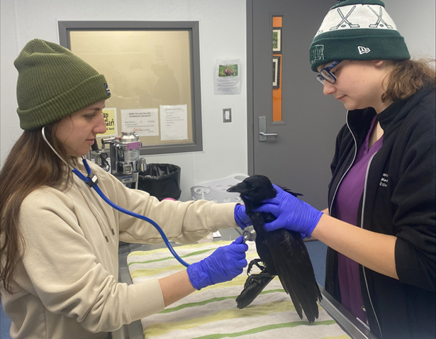
During winter break I volunteered to come back to school early and help in the clinic for a little under two weeks before classes resumed. While I was working in the clinic, we had a crow that was in our care. I was made primary on the case, which meant I was the first person to be contacted if something was going on or someone had questions and I was the main person who would guide medical decisions. I was very excited for the opportunity and challenge but also terrified that I wasn’t ready. Luckily, the secondary on the case was a third-year veterinary student and team leader that I had previously worked with, so I was excited and relieved to have their expertise and experiences to help with this case.
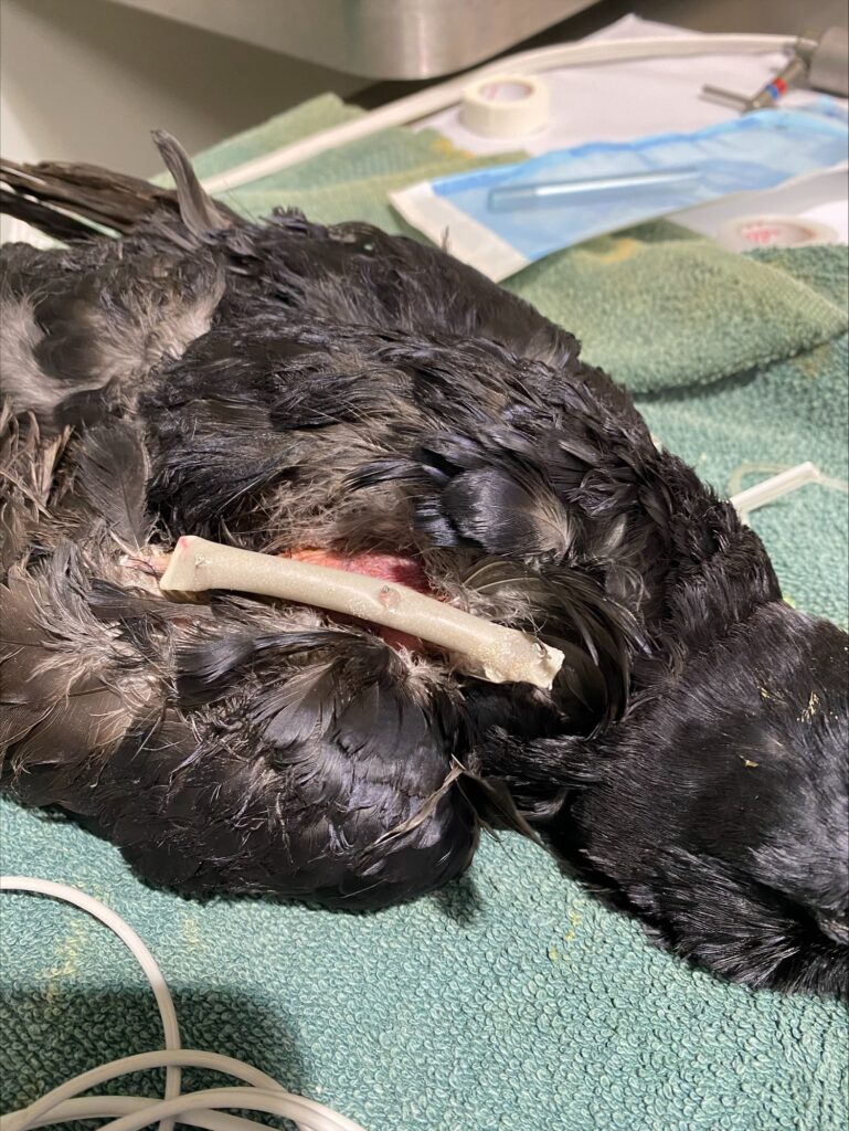
This was taken post surgery and the white bar you see is the external fixator.
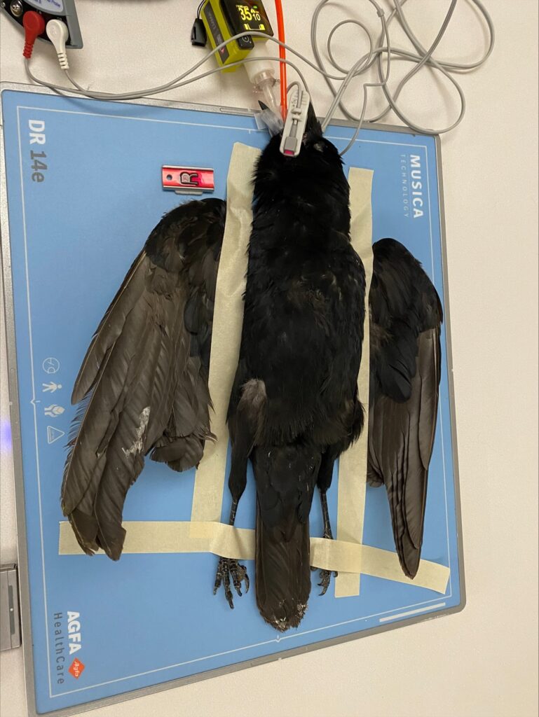
The crow came into the clinic in late December with a broken right humerus (a major bone in the wing). We were able to perform surgery to repair the break, which involved placing pins to hold the bone in correct alignment while it was healing. We put one inside the bone across the two fragments and then we had some on the outside of the wing to help stabilize everything. While the crow had his pins in his wing and the bone was healing, he couldn’t move it as well as he normally could. Therefore, we performed passive range of motion or physical therapy for the crow three times a week. This also allowed us to gauge his progress in healing. As the weeks went on, he was able to stretch his wing more and more, and eventually his bone was healed enough to remove the pins. We took new x-rays and confirmed that the bone had healed adequately, and we could start removing some of the pins.
First, we cut the connection between the pin inside his bone and the pins outside his bone. A few days later we were again performing his physical therapy but noticed that he had pulled out the pin that was inside his bone, all on his own. We once again took x-rays and confirmed that the bone was healed, and then proceeded with removing the rest of the pins. Crows are extremely smart and like shiny objects, so we think he noticed that the pin inside his bone was no longer attached to the external ones and was clever enough to pull it out on his own. Once all the pins were removed, we allowed our patient to continue to cage rest in a small space.
The patient getting radiographs taken.
One week later, just before we hoped to transfer our patient to a wildlife rehabilitator for some time in a flight cage, we noticed bruises present over his right wing. Radiographs confirmed that he had rebroken his wing and in the exact same spot that had just healed! We are not sure exactly how he re-fractured the wing, but he was quite active in his enclosure, and may have hit it firmly against the walls of the cage. We again took this patient to surgery to place a slightly different type of external skeletal fixator. We moved him to a new enclosure in his own room and made sure everything was as soft and padded as possible and began the healing process again.
We increased the amount of enrichment provided to the crow by means of food puzzles and various toys to try to keep him busy and occupied so his wing could heal. As I mentioned before, crows are very intelligent animals and when we are caring for them it is part of our job to make sure they are using their brains and are mentally stimulated. We have some great puzzles that we hide their food in, various objects such as bells and mirrors that we had in this crow’s enclosure for the duration of his stay, and a variety of food to keep his diet interesting.
We fed this crow a mix of dry and soaked dog and cat food, wet dog food, rolled oats, cracked corn, berries, hard-boiled eggs, and variously sized mice. We also did our best to be as hands-off as possible and handle the patient only when absolutely necessary, which greatly helps to reduce stress while they are in our care. Following the second surgery, we began to restart physical therapy. On his first physical therapy session we found fresh bruising yet again. This once again led to further x-rays, which showed he had broken the same bone in a new location.
Roxanne with past manager Bri and new manager Riley doing an exam on the crow.
The new fracture was closer to his elbow than the original fracture. At this point, it was the crow’s second fracture of the bone in our care and the third fracture of that same bone within two months. After much discussion, we made the hard decision to humanely euthanize him based on the severity of the newest fracture and its proximity to the elbow. With all the injuries, we couldn’t guarantee that the crow would regain range of motion in that wing and been able to fly comfortably once released. We were also worried there might have been a nutritional or metabolic abnormality that led to repeat fractures.
We did the best that we could for the crow while in our care, and unfortunately, while it was not the outcome we hoped for, it was the right decision and the most humane option for this patient. Although this case did not have a positive outcome, I was still appreciative of the learning opportunities it provided. Upon reflection on this case, I recognize how much I have grown as a veterinary student. I was able to work very closely with the faculty, residents, and the managers throughout the process. I was able to build relationships with all these amazing people who are involved in the clinic by working on this complicated case with them. My team lead/secondary was there to help answer my millions of questions and really increased my confidence level. The faculty were a great support system for the harder questions we had while working with this crow and were there to help us every step of the way.
Without the mentorship of my team leader and the faculty in the clinic, I would not have learned as much as I did, and I’m extremely grateful to them for helping me. Working on this case allowed me to learn about avian orthopedics, the process of dynamic destabilization (taking the pins out in stages), case management, and treatment decisions which I will be using throughout other cases during the rest of my career. I enjoyed being that first point of contact to always be up to date and involved in the case. It was very intimidating to shift my thinking of myself from a student to a veterinarian. Making that shift mentally allowed me to think more about the medical decisions we were making, and I felt that I was very involved and integrated into the case.
When I think about my experiences in the Wildlife Medical Clinic so far this year, it all centers on increasing my clinical skills and confidence, and this case was instrumental in that. I am very lucky that I’m already hands-on with the entire medical process as a first-year veterinary student, and I can’t wait to see how much more I learn, grow, and gain from working with all the amazing other students, faculty, residents, and animals that we interact with at the wildlife medical clinic.
What do Spiderman, Iron Man, Wonder Woman, and the Veterinary Student all have in common? They are all heroes. Ever since I was a child, I have dreamt of becoming a superhero. I pictured myself as this fearless, gallant character in a comic book. A bold superhero who saves the lives of animals and people. Now that comic book is coming to life.
Let us begin with our exposition. Some heroes acquire their abilities, while others are born with them. The poor sickly orphan, Peter Parker gained his superhuman strength and speed once he was bitten by a radioactive spider, then took on the heroic name of Spiderman. It is simply not that straightforward for us. Veterinary students are special as they must work super hard to become the heroes that they are today. It was their unconditional love for animals of all shapes and sizes that called them to this career. A career that is centered around mute patients who can’t communicate their problems, so you are left with detective work to discover solutions. The average veterinary student knew they wanted to become a veterinarian the day that they knew that they could use the power of science and medicine to save animals. An ordinary moment that is captured at the beginning of many veterinary students’ journeys through a baby picture is them wearing their elevated costume of scrubs covered by a white coat with the dynamic equipment known as the stethoscope wrapped around their neck sitting next to their beloved pet sidekick. Our costumes might not look as marvelous in comparison to Superman, the Flash, or Black Widow. However, once we put on our costumes, we feel invincible.
As a child, I would beg my mom to take me to the zoo almost every weekend because I was fascinated by all the unique zoo animals. My mom expressed to me that she knew that I would become a veterinarian when she simply saw the joy that animals would bring me.
The road to veterinary school is complex. Most students attend veterinary school after obtaining a bachelor’s degree from an undergraduate college. However, others go through a nontraditional path and return to academics once they discover that veterinary medicine is their true passion. To become a Doctor of Veterinary Medicine, it requires precision, devotion, and intensive training. The exposure to numerous specialties allows students to find their calling to work in specific fields such as companion, farm, equine, zoological, aquatic, and wildlife medicine. Our opportunities are endless. They all share one similarity: being a healer for animals. When I first stepped into the Wildlife Medical Clinic, I was mesmerized by the ability to save the lives of injured and orphaned wildlife. I knew that this was my calling. A future in saving nontraditional species such as zoological, aquatic, and wildlife animals.
Like all superheroes learn quickly, team dynamic is necessary to become successful. We cannot do this alone. This clinic is student-run with the managers directing over eighty students broken into six teams with bold, courageous team leaders guiding the team members. We are a community of heroes. This clinic operates 365 days with the help of undergraduate students, veterinary students, house officers, and faculty members devoting countless hours. Outside of an intensive rigorous curriculum, students volunteer their time when they are not in class to provide the best care to patients. Many students dedicate their time to learning more about wildlife medicine, husbandry, nutritional needs, etc. Overall, we are working together to not only save lives but make a difference in this world. Like all superheroes, we learn the impact of allies. We depend on each other.
The climax is the moment that challenges every superhero and leaves them with difficult choices to make. During the summer of 2023, every day was filled with adventure from feeding orphans to triaging patients to providing medical treatment. At the end of a very long day, we were notified that an injured five-day-old white-tailed deer was waiting in the front lobby to be admitted to the clinic. This patient was in rough shape and presented as weak, non-ambulatory, and almost lifeless. The team quickly sprang into action and used whatever power that they had left and put it into this fawn. After providing intensive medical care, the outcome was not what they hoped and worked for. We spend most of our days in the clinic staying positive and living through joyful moments. Even so, we are sometimes faced with the realization that we can’t save them all. We are reminded that even for the patients that we lose, so many others are given another chance at life and released back into the wild through incredible care such as a red-tailed hawk with an ulnar fracture, a bald eagle with lead toxicosis, or a sickly red fox with mange. Even when we are faced with a difficult decision, supporting an animal through its toughest moments is the most courageous thing that we can do. That is the true power of our field.
Our journey through the Wildlife Medical Clinic has taught us to be advocates for all animals. Our work is not only done in an examination room. We can inspire the world to strive for lifelong education and preserve the environment through conservation efforts. We are in this field to be heroes for our patients. We are here to make a difference in this world. My comic storyline is not over. I have many unwritten pages awaiting me. Patients for me to learn and grow from. The Wildlife Medical Clinic will always be a monumental moment in my journey as a veterinary student.
Not all heroes wear capes – but these veterinary students do.
This article was written by Hassan, Class of 2025 DVM Candidate
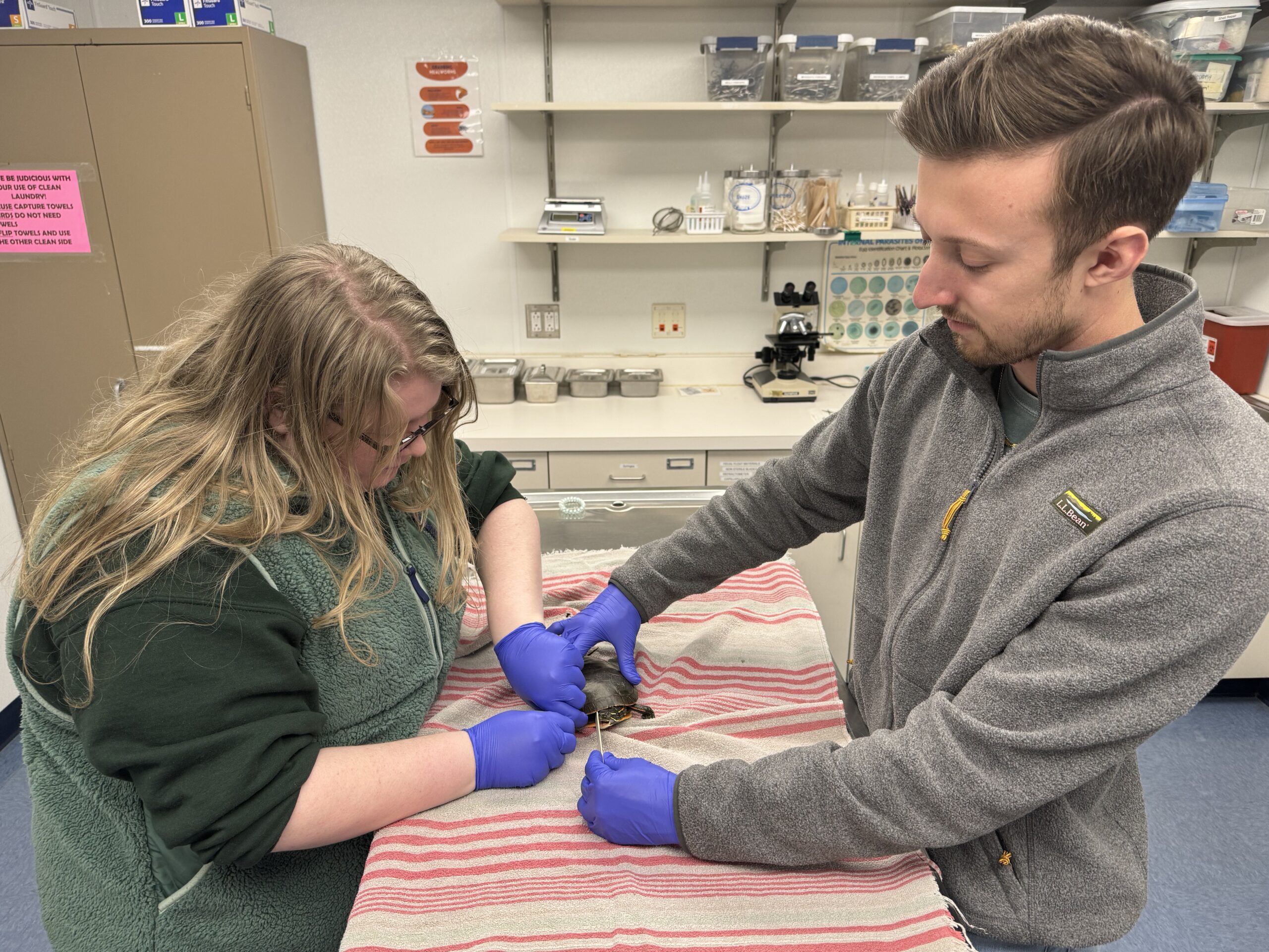
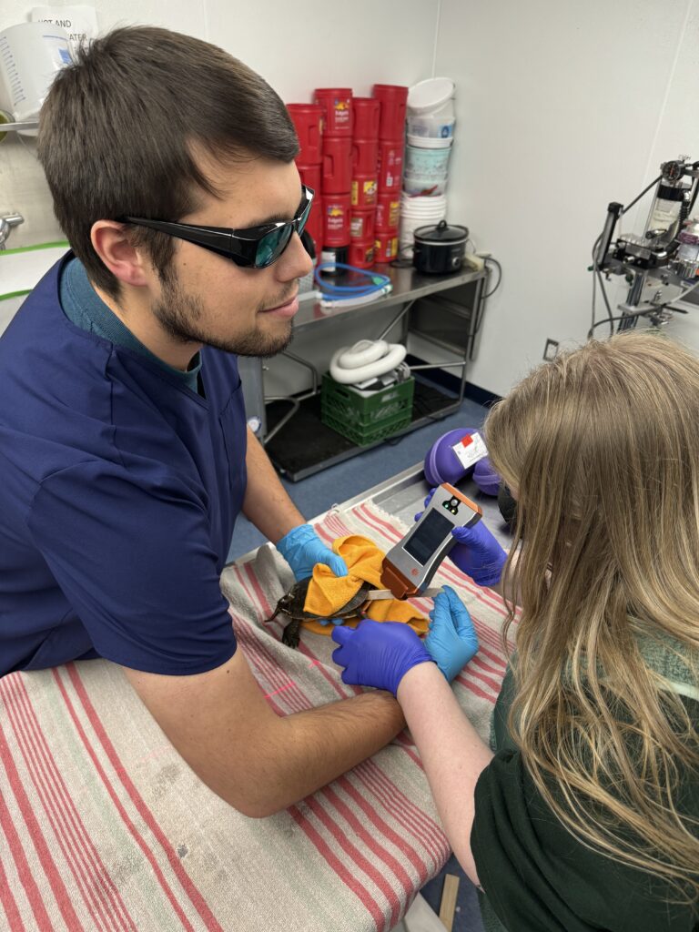
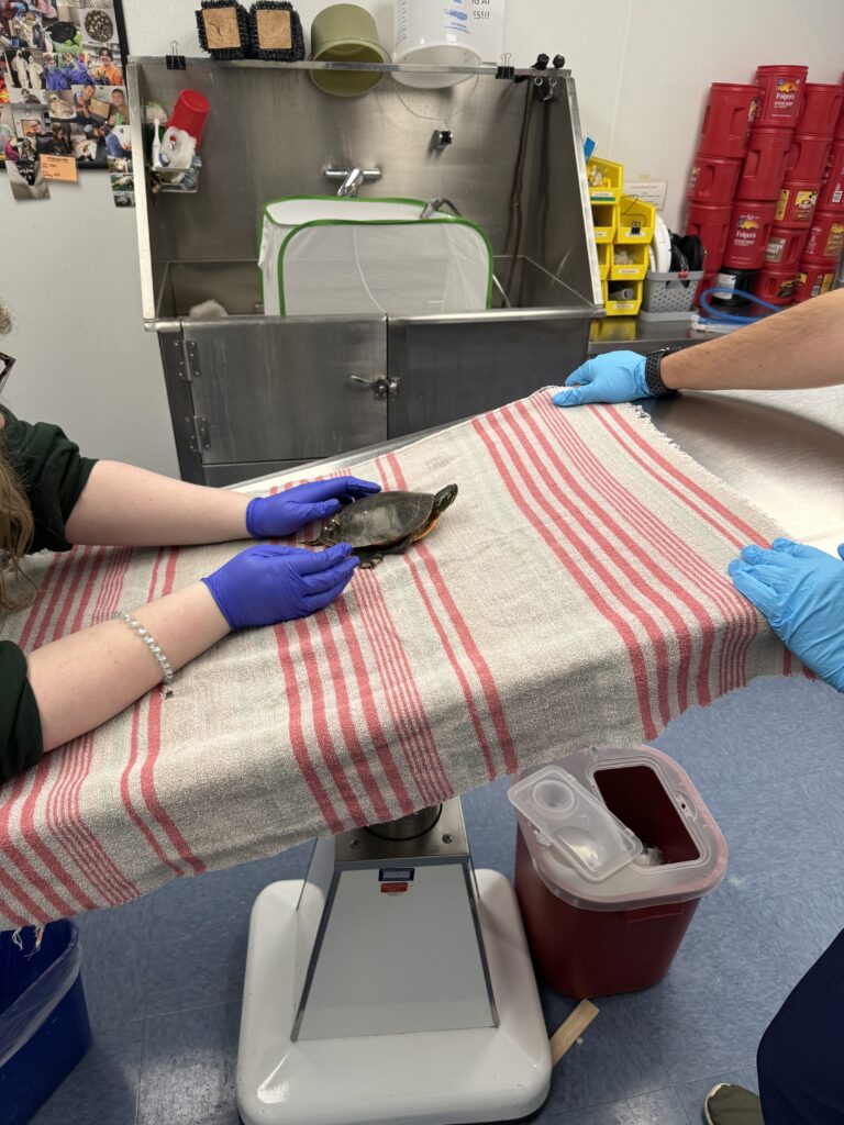
At the end of August, an adult male Midland Painted Turtle presented to the Wildlife Medical Clinic from a rehabilitator after it was suspected to have been hit by a car. This patient had a fairly severe fracture of his carapace (top of shell) and required extensive treatment to repair it. While receiving these treatments, the turtle developed neuropathy (nerve damage) in his right front leg. This problem caused him to incorrectly place his foot when walking. To correct the placement, we started him on a routine of PROM, laser therapy, and physical therapy three times a week.
PROM stands for passive range of motion and is performed by slowly and repetitively stretching the joint. The goal of PROM is to increase the stretch and movement of the structures that surround the joint. For this specific patient, PROM consists of extending the carpus (wrist) & phalanges (fingers) as far as possible, three times for thirty second intervals.
We are using laser therapy to increase healing and blood flow to the limb as well as to decrease pain, inflammation, and swelling. This patient’s front leg has been inconsistently swollen since the nerve damage took place. With the laser therapy, our goal is to decrease the swelling and promote faster healing. We perform the lasering from the top of the radius/ulna (area between elbow and wrist of front leg) to the tips of the phalanges (fingers) to achieve this goal.
The final part of this patient’s therapy plan is physical therapy. When developing a physical therapy plan, it is important to focus on a specific area while building up strength and correcting incorrect habits that have developed. We complete this through repetitive exercises. Our turtle’s physical therapy plan involves walking up a ramp six to seven times per session, while a volunteer places their fingers behind his limbs to help him properly place his foot on the ramp.
Overall, therapy in reptiles is like other animal species and is tailored to the patient’s specific needs. This patient has been showing great improvement with this therapy plan and we plan to continue until the spring weather reaches Illinois, which is when he will be able to be released back into the wild.
This article was written by 2nd year veterinary student Becky.
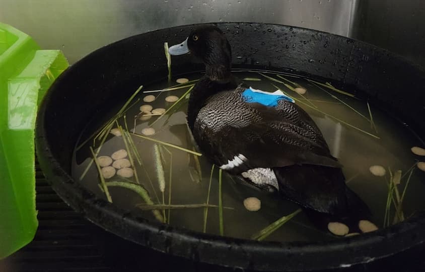
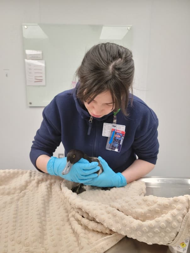
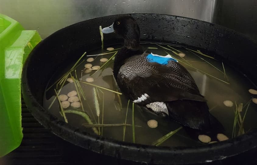
On February 28, 2023, an adult male lesser scaup presented to the Wildlife Medical Clinic. This is a type of duck, and they are sexually dimorphic, meaning the males and females do not look the same. Males have black and white feathers and a blue bill, whereas the females have more brownish colored feathering and a white patch next to their black bill. They typically migrate from Central America, Mexico, the Caribbean, and the southern United States to the Northwest United States and Canada. The spring migration begins in February, so we believe that this patient was passing through Illinois on his way north. Our initial goals were to rehabilitate him as quickly as we could so we could reunite him with his flock on their way north to their summer home. Lesser scaups are diving ducks and like to spend most of their time on the water. They will dive in waters less than 16 feet deep, using their feet to propel them as they forage for prey.
During his physical exam, we appreciated an injury in the right shoulder. We suspected that the injury was due to a traumatic event. The exam showed no other abnormalities, which meant the patient was otherwise healthy. To stabilize our patient and prevent further injury until one of our veterinarians could examine him, we placed a bandage to secure the wing to the body. We were hopeful that the injury was one that could be healed and eventually allow our patient to regain full flying capabilities.
In a few days, the patient was anesthetized to obtain radiographs (x-rays), and sadly we found that the injury was severe and could not be corrected well enough for this bird to ever regain normal flight.
This was a difficult case that I struggled with, because of course I became interested in veterinary medicine to rehabilitate injured animals. My experience prior to working in the Wildlife Medical Clinic had been primarily with domestic animals, but the goals are completely different when working with wildlife. When working with domestic animals, our goal may simply be to provide a comfortable quality of life in the care of their owners. Wildlife, however, need to be indistinguishable from other members of their species, as they need to be functioning optimally to ensure their survival in the wild. After we release them, wildlife need to migrate, find food, evade predators, mate, and raise offspring. We can’t provide them with life-long medications, or have them return later in life for recheck exams. Any animal that cannot perform these functions would not survive in the wild, and many chronically injured wild animals would not be suitable for a life in captivity. These high standards are what makes wildlife rehabilitation so difficult.
At the end of our journey with our patient, we were still able to help end his pain and suffering. As veterinary students and veterinarians, we are responsible for making the most informed decisions for our patients, no matter how difficult they may be. I am grateful that I learned this valuable lesson as a student in the Wildlife Medical Clinic. While the outcome wasn’t what I hoped for, it was still the best outcome for this individual and helped me to learn and grow in my journey to becoming a veterinarian.
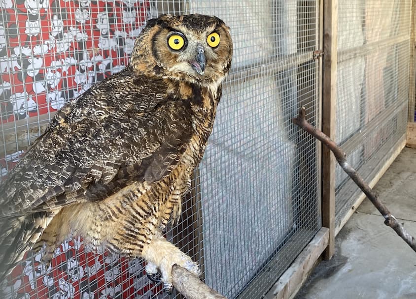
The great horned owl, Bubo virginianus, is a common patient seen at the Wildlife Medical Clinic. This species is found throughout most of North America and is easily identified by its “horns” which are actually tufts of feathers. Great horned owls are permanent residents of Illinois and can be found in a variety of habitats. This species is also known for its stereotypical “hoo-h’HOO hoo hoo” call, which is commonly heard in movies and on television shows.
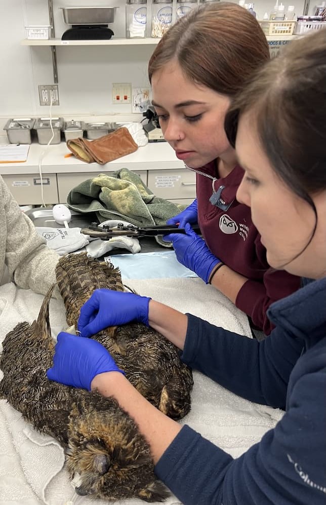
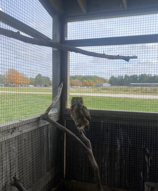
In the middle of August, a juvenile great horned owl was brought to the Wildlife Medical Clinic by a good Samaritan. The owl had been in the finders yard for a few days and was seen taking short flights but would fall out of the trees upon landing. The finder also noticed that the young bird was not using its right leg at all, and it seemed to be dangling from the joint. During the physical exam, the patient was found to have a proximal fracture of the right femur. The patient was also found to have external parasites, blood parasites, anemia, and was extremely underweight. Shortly after the initial exam, the patient had radiographs performed to confirm the fracture location and to evaluate whether surgery would be an option to repair the fracture. Upon review of the radiographs, it was determined that fracture repair was possible and had great potential to be successful, so the patient was taken straight to surgery.
During the surgery pins were placed in the bone to stabilize the fracture and allow for the fractured pieces to be joined back together. Over the next six and a half weeks the patient was placed on cage rest while the bone healed. During this time, the patient was placed on antibiotics to help prevent infection, as well as medications to control pain and decrease inflammation or swelling. The patient was also treated for its parasites, which in turn treated the anemia the patient presented with. This period of rest also allowed the patient to gain weight and return to a healthy body condition. The patient’s surgical site was monitored daily for signs of healing and was kept clean by the volunteers. During the last week of cage rest, radiographs were repeated and confirmed that the patient’s fracture was healing properly and the surgical implant holding the bones in the correct position was ready to be destabilized. Destabilization essentially allows for the bone to slowly get used to supporting the weight of the patient.
When the pins were removed there was slightly more exudate than what would normally be expected. The material was cultured and sent to the Veterinary Diagnostic Laboratory at the University of Illinois to be examined by pathologists. The sample came back as an antibiotic resistant bacterium, staphylococcus epidermis. The bacteria was tested against different antibiotics until one was found to be effective against it. This type of infection is known as osteomyelitis, or a bone infection. Osteomyelitis happens infrequently but is associated with surgical repairs using pins in raptors. The patient was put on a course of twice daily antibiotics, and pain medications.
During this time, the owl was also moved to a larger enclosure in our flight cage, for further monitoring. This allows the doctors to confirm that the bone is continuing to heal properly and that the patient can use the limb correctly. This also allows for the patient to get used to using its leg and rebuild any muscle that may have deteriorated during the healing process. This is especially important because owls are birds of prey, which use their legs and talons to capture the food they eat. Owls also nest and perch in trees, which allow them to stalk their prey and stay safe from predators. Proper limb function is needed to keep the bird steady in the tree, and to catch their prey silently.
After three weeks of antibiotics and pain management the medications were discontinued, and the patient was monitored for any signs of pain, discomfort, or improper limb use. During this observational period the patient showed no clinical signs of infection or pain. Radiographs were also repeated and showed no evidence of damage to the bone from the infection. Upon further examination the patient was deemed ready for transfer to a rehabber to build up strength and sharpen its skills before being released to the wild. This is an important success story as Osteomyelitis is a rare surgical complication that can lead to the downfall of a patient. Luckily, the infection was caught early, and the patient responded well to the course of antibiotics giving him a chance to return to the wild, where he belongs!
Red-tailed hawks (Buteo jamaicensis) are one of the most common hawks in North America, with a range that spans from Alaska to the tropical rainforests of Central America. You can often find them perching on fences and telephone poles, surveying their territory. While you may not have heard their calls in real life, you’ve definitely heard them in a movie theater. The call of a red-tailed hawk is the standard sound effect for most birds of prey in movies, especially bald eagles.
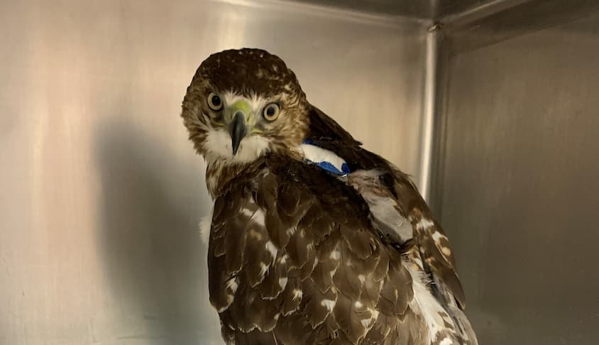
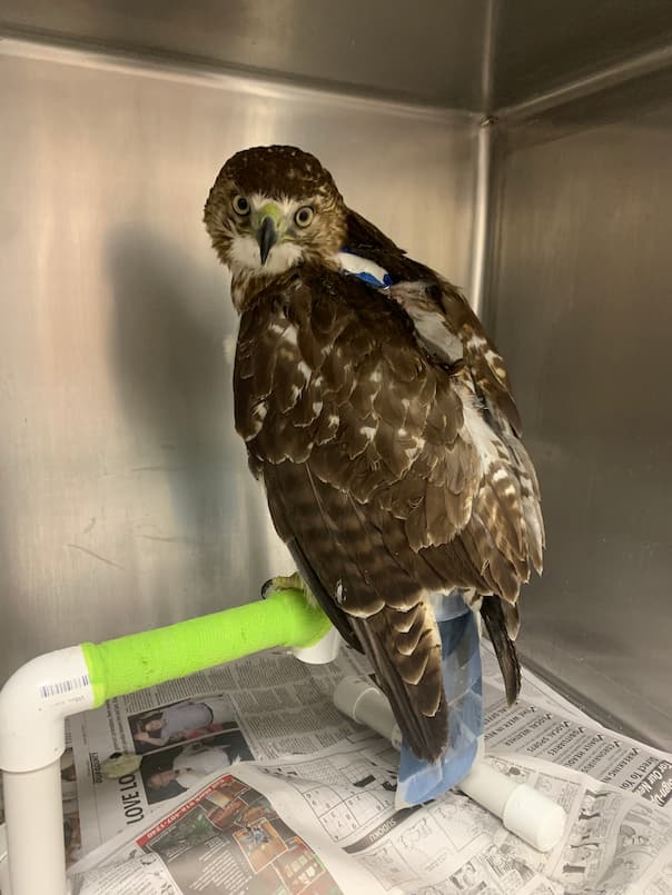
Here in Illinois, red-tailed hawks are year-round residents, choosing to stay put once winter comes. Back in January of 2023, an adult red-tailed hawk was brought to the clinic by a good Samaritan after being found on the side of a road in Champaign. The finder was able to catch and carry the hawk by wrapping it in a towel they had found on the road next to it. After bringing the hawk to the clinic, we were able to perform a triage exam. The hawk was dull and squinting, signs that can point to pain. We placed a raptor hood, a useful tool that covers a hawk’s eyes, calming them down to allow us to safely perform a thorough exam. This helped reduce his stress while we continued our exam, feeling the joints and bones of the wings and legs. The hawk’s right wing had an ulnar fracture, and several wounds were found. Additionally, we were able to feel bony changes on the left leg, indicating that the femur of the leg had fractured previously and was healing.
To get a better look at the bony changes we had felt in the right wing and left leg, we took radiographs of the hawk. The radiographs confirmed the ulnar fracture, also revealing that it had multiple bone fragments instead of a simple, clean break. The femur of the left leg was fractured in two locations but had since rejoined together and was healing well. The leg fracture was approximately a month older than the wing fracture, meaning this hawk had experienced two traumatic events in a short period of time and still had a zest for life!
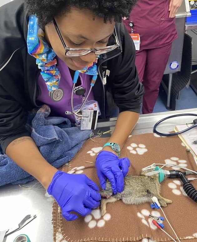
Passive range of motion, often abbreviated as PROM, is a form of physical therapy in avian patients in which the wing is gently and repeatedly stretched to its normal limits while the patient is anesthetized. The angles for the maximum extension of the elbow and wrist are measured before and after performing PROM, allowing us to track how well the patient can spread their wings. Birds need a very specific range of motion to fly effectively, so it is important to maintain that range of motion after an orthopedic injury like this hawk had experienced. Regular PROM also provides the students working in the clinic plenty of opportunities to practice valuable veterinary skills such as anesthesia monitoring and placing wing wraps.
Over the course of eight weeks, we were able to see steady improvement in this patient. The final radiographic examination revealed that the various pieces of bone had bridged, and the wing was stable. This hawk was finally cleared for transfer to a rehabber, allowing it time to practice and improve its flying skills again before being released to the wild.