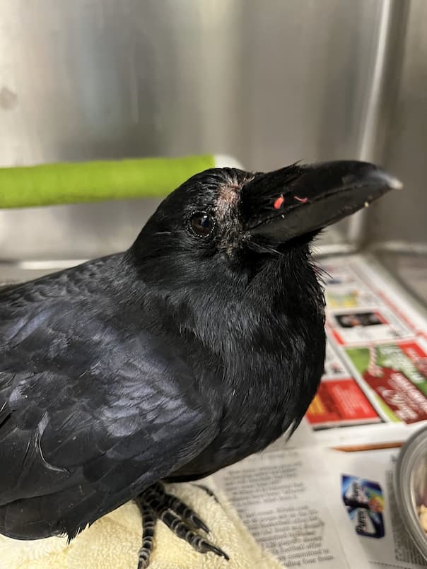
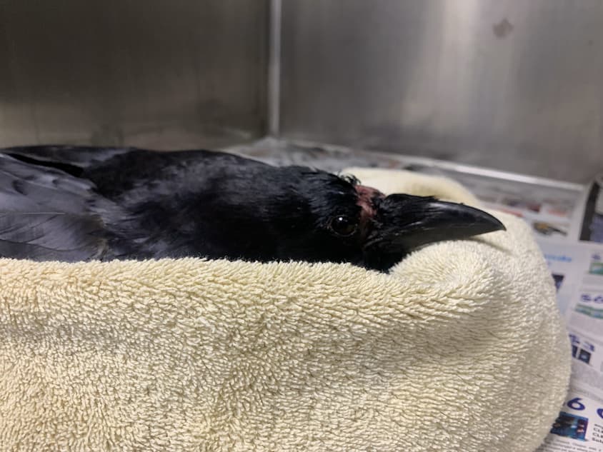
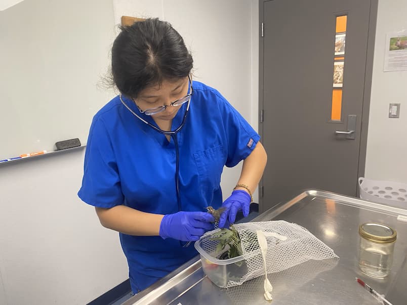
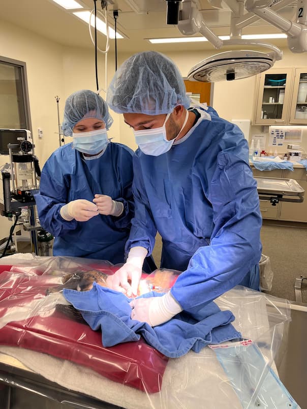
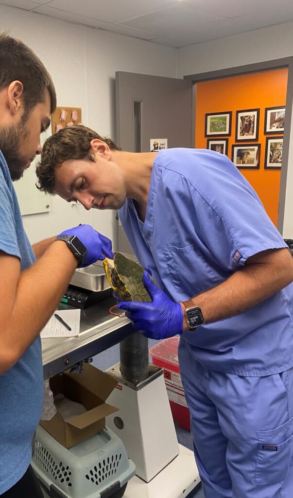
American crow (Corvus brachyrhynchos), one of the most intelligent bird species in North America. They are very social birds and sleep in a communal roost together in the winter! They thrive by working collectively and using their intelligence to catch or steal prey and other food items. Yet, this unique species has faced the threat of West Nile Virus since 1999 when the outbreak began in New York City. According to the data, this disease has caused a 45% regional decline in crow populations.
West Nile Virus (WNV) is a vector-borne disease that is spread by mosquitoes. In a temperate climate, WNV is prevalent in late summer and early fall. It can infect a wide range of species, from birds to humans. Mosquitoes play a critical role in the transmission, as they are the vector. Meaning they are the intermediary. They get the virus from the blood of one host, for example an infected bird, and then when they bite another species like a human, they pass the disease through the bite. Transmission has also been documented from bird-to-bird. This usually occurs when a bird consumes another bird that is infected with WNV or when water tainted with feces and saliva from an infected bird is drank.
Over 250 bird species have been documented as WNV-positive cases. Within these species, the corvids (crow, raven, and jay) have the highest susceptibility to WNV in North America. Many studies have investigated this phenomenon and found correlations between human WNV cases, mosquito transmission pathways, and crow death from WNV. Rapid urbanization significantly impacted the speed of virus transmission in wildlife.
West Nile Virus in birds primarily attacks the central nervous system. Therefore, some of the symptoms an infected bird displays include a wide range of neurologic signs, such as a head tilt, lethargy, weakness, and loss of coordination. Many cases that come across the Wildlife Medical Clinic also show a great degree of emaciation, where patients are significantly underweight and are unable to fly. Once the crow is infected with the virus, it almost always indicates a grave prognosis. Studies have shown that most infected crows die within three weeks of contracting the virus. And testing for the disease will likely occur post-mortem through tissue samples from the heart, brain, and kidneys or via oral swabs.
The success rate of recovery is incredibly low in crows, since no known established treatments are available to clear the viral infection. Most cases rely on the animal to clear the virus and recover with supportive care. And the mortality rate can be as high as 100%. However, some breakthrough discoveries were made in 2022 by the Cornell College of Veterinary Medicine – where their wildlife experts successfully treated five infected crows with fluids, anti-inflammatory drugs, anti-fungal drugs, and anti-parasitic drugs. This treatment successfully allowed these wildlife experts to release the five American crows that were previously ill with WNV. The discovery was linked to potential immune system adaptation in the crow or the shifting of the virus’s behavior. While this treatment was successful, cases in general still pose a grave prognosis for crows and research continues in order to better understand this disease dynamic. We are hopeful that a treatment will become available in the future that will help our Crow friends fight off this deadly virus.
Written by YiYing, Class of 2026
References
Wheeler, S.S., Taff, C.C., Reisen, W.K. et al. Mosquito blood-feeding patterns and nesting behavior of American crows, an amplifying host of West Nile virus. Parasites Vectors 14, 331 (2021). https://doi.org/10.1186/s13071-021-04827-x
Yaremych SA, Warner RE, Mankin PC, Brawn JD, Raim A, Novak R. West Nile virus and high death rate in American crows. Emerg Infect Dis. 2004 ;10(4):709-11. doi: 10.3201/eid1004.030499. PMID: 15200865; PMCID: PMC3323091.
Many of you have found an injured or orphaned animal and brought it to us, but do you know what happens after you drop it off? Come with us as we walk you through everything that happens when wildlife visits the Wildlife Medical Clinic here at the University of Illinois Urbana-Champaign!
When you bring an animal to the front desk, they send out a page to all the volunteers in the clinic. Countless phones throughout the college light up with “Wildlife Medical Clinic Receiving:” and the patient’s species, if known. The volunteer team assigned to triage that day then springs into action, heading to the clinic to bring the animal back and begin the triage process. It is very important that a detailed history is given on the animal’s intake sheet, as this helps the triage team determine appropriate steps going forward. Certain species that are susceptible to Canine Distemper Virus, such as raccoons, bobcats, and coyotes, should be left in your car until the triage team arrives to transport them, as the lobby is a shared space amongst many animals also susceptible to this disease. It is always worth calling prior to bringing in an animal for best practices and safety advice from our volunteers.
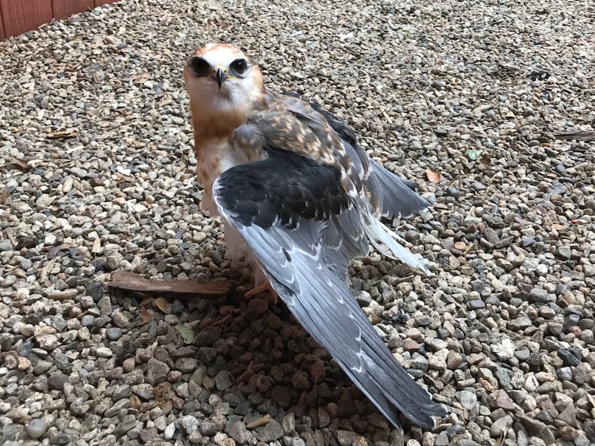
A white-tailed kite displays a wing droop.
Once the animal has been taken back to the designated triage room within the clinic, volunteers begin with a hands-off exam. A lot of important information can be gained simply by looking at the animal before it has been touched by the volunteers. Is it standing? Are there any visible wounds? How does it react to the presence of humans? Even something as simple as how a patient is holding themselves can tell us where an injury may potentially be. Take the image of a white-tailed kite, a species found in California, Texas, and parts of Mexico. The way the kite is holding its wing slightly out and away from itself is called a “wing droop,” which can be indicative of an injury to the wing or the flight apparatus that physically supports the wing.
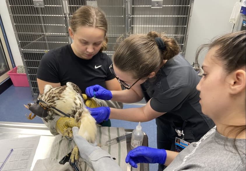
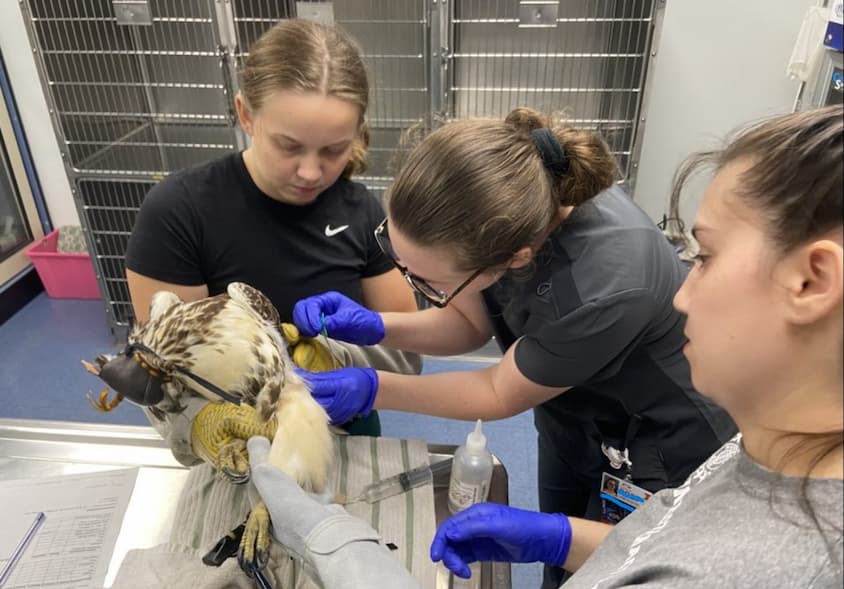
Students restrain a red-tailed hawk wearing a falconry hood for fluid administration.
When the hands-off exam is complete, it is time for volunteers to restrain the patient for a physical exam. Proper restraint is important for the safety of both the volunteers and the patient. There are two types of restraint used: physical and chemical. Physical restraint is used for smaller patients and raptors and involves using specialized gloves that protect human hands from bites, scratches, and the vice-like grip of a raptor’s talons. All volunteers are trained in how to properly hold different species so that they cannot injure themselves or others. Chemical restraint, or the use of sedative medications, is used for carnivores, meso-omnivores, and other species that pose a more significant risk to volunteer safety when awake, as well as patients that are exhibiting signs of stress and would benefit from the anxiety-reducing effects the medications provide. A falconry hood is an alternative option that can be used in raptor patients. This leather mask completely covers the raptor’s eyes, keeping them calm and comfortable during what would otherwise be a very stressful time.
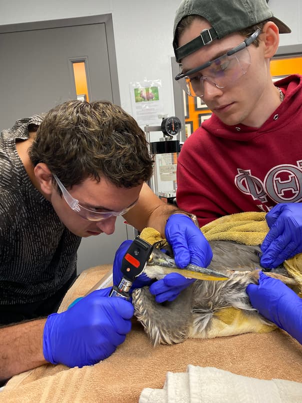
A student intern and manager perform an ophthalmic exam on a blue heron.
After the patient is properly restrained, the next step is to take vitals. Basic vitals include a heart rate, respiratory rate, and temperature. These provide insight into the patient’s status, allowing the triage team to quickly make decisions. The goal of a triage exam is to stabilize the patient so that procedures and diagnostics (such as bloodwork or radiographs) can be performed in the future. Stability can change rapidly, but monitoring vitals gives the triage team the information they need to provide the medications and treatments that can improve stability and support the patient.
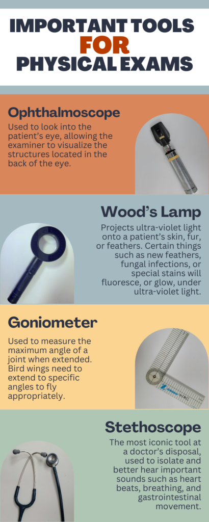
Now that vitals have been recorded, it is time for the physical exam. Every volunteer has a different approach they utilize to complete an exam, but every exam must involve careful observation of the presenting animal’s entire body. A good physical exam can prevent unneeded procedures, reducing the time an animal needs to be handled and thereby decreasing the animal’s overall stress. Examiners use a combination of sight, touch, smell, and hearing as well as various tools to gain a complete picture of the animal’s status.
The ultimate goal of wildlife rehabilitation is to release the animal back into the wild. For this to happen, the animal needs to truly function as a wild animal; they must be able to successfully find or catch food, move and maneuver in a typical way, demonstrate appropriate behaviors including fear of humans, and possess all of the physical characteristics they need to regulate themselves, among other standards. If the triage team determines that the animal will not be able to meet those standards, even after treatment and support, then the animal is recommended for euthanasia. The purpose of euthanasia is to end suffering, but wildlife practitioners have the unique dilemma of also determining the possibility of future suffering. Most of our wildlife patients are not going to return to the clinic when complications of their problems arise or worsen post-release. To release a wild animal knowing that it is at a disadvantage in the functions it needs to survive is to cause suffering. The triage team presents the case history, their exam findings, and their recommendation to a licensed veterinarian. The veterinarian then either approves the recommendation or offers advice on treatment options that may work.
If the problems the patient is experiencing are treatable, then the triage team gets to work providing supportive therapies. These often include fluid therapy (most wildlife patients present with some level of dehydration), anti-inflammatory and pain medications to keep injured patients comfortable, and other treatments more specific to the patient’s case.
Since we are a student run facility, we depend on you and the rest of our community to bring these patients to us when they need help. Without your support, we would not be able to do what we do. We hope you enjoyed this special look into our triage exam process and the work we do here at the Wildlife Medical Clinic.
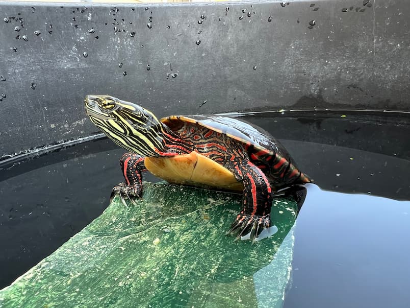
The Wildlife Medical Clinic commonly receives turtles in need of shell repairs. Recently, an injured adult Midland painted turtle (Chrysemys picta) was admitted to the clinic for “having a cracked shell.” Chrysemys picta are the most widespread native turtles of North America. They are normally found in slow moving freshwater. These turtles do not migrate and will stay in a single area their entire lives. Due to their unique markings and vibrant red and yellow coloring, these turtles are easily identifiable. These markings can be seen on the lower portion of the shell which is called the plastron and the upper jaw. This specific turtle was identified as a male based on the length of the nails, and the placement of the cloaca as these features differ between males and females, as well as overall size of the individual.
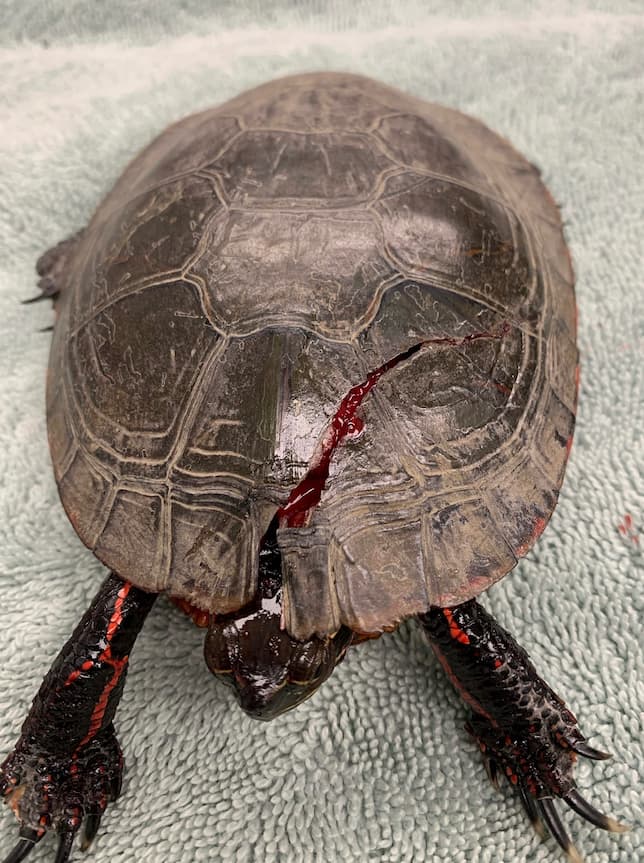
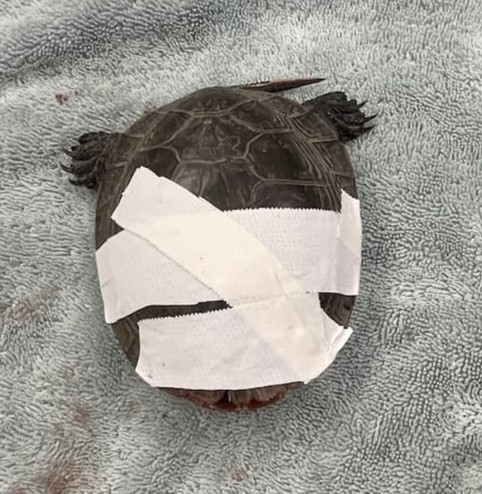
During the initial exam, a large fracture was found on the carapace or the top portion of the shell. The fracture started from the cervical scute which is the middle of the shell near the turtles head and crossed over to the left bridge of the shell, which is basically the side of the turtle, but the middle where the carapace and plastron are connected. Thankfully, no crack was seen on the plastron, so this turtle had a better chance of recovering from its injuries with its shell completely intact. However, it was not evident on the examination whether the trauma extended into the body cavity affecting his lungs and other organs. Although the patient was actively trying to escape, the team noticed that his eyes were closed the entire time he was being handled. This indicated to the team that the animal was in pain, which was to be expected given the extent of his injuries. The exam was paused so the appropriate pain medication could be given to make the patient more comfortable during the exam. Once the team was sure the medication was working, they continued with the triage exam. No other injuries or ailments were noted during the exam, meaning the turtle seemed to be healthy minus the pressing issue of this large shell fracture. His wounds were gently and thoroughly cleaned before the triage team administered an injectable anti-inflammatory medication and an antibiotic, in addition to the pain medication he had already received. This course of treatment was given to help reduce swelling and prevent infection in the wounds. Tape was then placed on the shell to stabilize the fracture until radiographs could be performed.
When it came time to do radiographs, pain medication was given prior to the turtle being handled to ensure he was comfortable during the procedure. The tape was carefully removed, no signs of infection were seen, and the carapace had already begun healing. The radiographs showed the fracture did not go into the body cavity, which meant no organs were damaged by the initial trauma. This was great news and meant our patient was a good candidate for continued shell repair! When doing a shell repair on any turtle, there are several approaches that can be done to have successful results. For this specific case, the shell was already starting to heal, so the team decided to go with the most common approach of placing cerclage wire into the shell. Cerclage is a type of flexible surgical stainless-steel wire used to stabilize bone fractures. Two small holes were drilled on each side of the cervical scute where the fracture initially started. Cerclage wire was then placed through the holes and tightened so the carapace fracture was closed and even on each side. This concluded the procedure.
Now we just watch and wait, giving the shell time to heal. Even with a successful procedure, complications can still arise. In this turtle’s case, he had been dry docked or not allowed to swim in water prior to the shell repair and this paired with the combination of drugs and limited amount of fluids given, made him slightly dehydrated. Thankfully, after the repair, the patient was able to be fully submerged in water once again and was given additional subcutaneous fluids which helped to rehydrate him and continue moving him forwards on his road to recovery. In the past, we have seen turtle patients refusing to eat after shell repairs. Inflammation and infection can cause patients to lose their appetite, so we will monitor him and his nutrient intake carefully to hopefully prevent this issue.
Due to the timing of this patient’s arrival, and the length of time a shell fracture takes to heal, he will be with us long term, staying over winter in the clinic. Thanks to our amazing staff and volunteers he is on the road to recovery and will hopefully be released in the Spring of 2024 after his wounds are fully healed.
This article was written by Jacquelyn Merx, Class of 2026
Introducing our newest house officers at the Wildlife Clinic: Dr. Erica Bender and Dr. David Minich! We look forward to seeing you around the clinic!
We asked them some questions to get to know each of them a little better.
Dr. David Minich, DVM, MAS, CertAqV
Where are you from? What vet school did you go to (and graduation year)?
I am originally from Cincinnati, Ohio. I attended veterinary school at The Ohio State University and graduated in 2021.
Why did you decide to pursue your residency here at the WMC?
The Illinois Zoological and Aquatic Animal Residency provides extensive exposure and training in the medical care of zoo-housed wild animals, native wildlife, and companion zoological species. These opportunities will allow me to develop my skills as a veterinarian and provide the highest level of care in my future career.
What are you most excited about working here at the WMC?
I am very excited to work alongside students on a wide variety of cases and patients, with the ultimate goal of providing the best care possible to native wildlife while supporting the education of veterinary students.
What is your favorite wildlife animal to work with?
I am particularly fond of both owls and bears, but I truly enjoy everything – it’s the diversity of species that drew me to this field!
What are your future career aspirations?
After completing my residency, I aspire to become board-certified in zoological and aquatic animal medicine and pursue a career in clinical medicine at a traditional zoo/aquarium, while also conducting research to further the knowledge and medical care of zoo-housed and free-ranging wild animals.
Anything else you would like to share?
I recently traveled to Laos and volunteered at a major wildlife rescue, rehabilitation, and release center. I hope to incorporate field-work such as this into my future career to help support in-situ conservation initiatives.
Dr. Erica Bender DVM
Where are you from? What vet school did you go to (and graduation year)?
I am originally from the San Francisco Bay Area in California. I attended vet school here, at The University of Illinois, and graduated in 2022!
Why did you decide to pursue your residency here at the WMC?
I am doing my specialty internship here, rotating between the WMC, our Zoo Medicine service, and our Zoo Ambulatory service. When I was a vet student, I volunteered and was one of the managers of the WMC and it has such a large part of my heart. I am super excited to be back working with all of our amazing faculty doctors and students, while getting to learn more about treating all the non-traditional species we see.
What are you most excited about working here at the WMC?
I am most excited about working with students and getting to be there as they develop their clinical skills and knowledge then apply them to treat our native wildlife species. Seeing their passion and the effort they put into treating our patients is always inspiring. And raptors, I’m very excited to work with raptors again!
What is your favorite wildlife animal to work with?
I spent a lot of time volunteering at The Marine Mammal Center in Sausalito, CA, so I’m a bit partial to working with California Sea Lions and Northern Elephant Seals. However, of our native Illinois species, my favorite species to work with are the red-tailed hawks.
What are your future career aspirations?
I want to be a zoo doctor when I grow up! I love the diversity of cases and the teamwork involved in treating animals in a traditional zoo setting.
Every year during the blazing hot summer, the Wildlife Medical Clinic welcomes its busiest season with a team of students eager to learn and ready to experience one-of-a-kind clinical experiences. This year it was four vet students from both first- and second-year classes joining the team and working alongside the house officers, student managers, and rotating clinical-year students. Their primary responsibility this summer was to support and facilitate treatments, triage patients, and discuss medical treatment plans with the students and faculty members. The clinic provides a unique opportunity to allow the student to sharpen their clinical skills and develop a deeper understanding of wildlife medicine and management. Summer interns are part of the foundation that ensures the clinic’s function. As we are gearing up to enter the fall semester at the end of August, let us celebrate all the hard work put into all the patients and the clinic by our outstanding summer interns. Below are some moments and memories shared by our 2023 summer intern team.
“One moment that really sticks out to me is when the team was able to release a red-tailed hawk that had been at the clinic for over a month. This bird had a wing fracture, which was surgically corrected, allowing for appropriate flight and the ability to be released back into the wild. That patient was transferred to a rehabilitation facility for further exercise and care, but it was extremely rewarding to see an animal make it through treatment successfully!”
– Jacob Dalen, Class of 2026
“We have had a lot of fawns (baby white-tailed deer) come through this summer, and this is a species that imprints easily on humans, so rehabilitation can be challenging. We don’t want them to get used to people, but they also need to be fed up to 5 times per day, so we had to follow strict protocols when taking care of them. I think my favorite moment this summer was loading five of the fawns into a van to go off to the licensed wildlife rehabilitator because it meant that we did our job well and gave them a chance at returning to the wild. But overall, I treasure just getting to work with the faculty, student managers, and other summer interns – we’ve spent countless hours together, but they make the long days go by fast!”
– Sarah Hollander, Class of 2026
“Working with all my amazing colleagues, learning from our supportive and knowledgeable faculties, and going through all the ups and downs together as a team.”
– YiYing Tung, Class of 2026
“The moment that I would walk into the clinic, I knew that I would leave the day with unforgettable, valuable memories. It has truly been a summer filled with hard work, laughter, and personal growth. I am so grateful to have had the honor of working with our incredible managers, ambitious house officers, driven clinical interns, and supportive faculty. I will always cherish the experiences and opportunities from this summer as it will play an important role in shaping my future as a veterinary professional.”
– Hassan Hanna, Class of 2025
Besides our summer interns, a group of students also played a critical role in the WMC over the summer. They are the members of our orphan feeding team, composed of 10-15 undergraduate and graduate students. Summer is peak baby season; The Wildlife Medical Clinic admits many orphaned patients. All of them need constant nutritional support, and without the orphan feeding team, we would not have been able to keep up with the needs of these patients. These students are crucial in the success of these orphan patients being transferred to a licensed wildlife rehabilitator for continued care. We want to share our great appreciation to all team members; the clinic is grateful to have such a fantastic team supporting the clinic’s operation and taking care of our orphan patients. Here are some memories shared by our feeding teams.
“This summer, in the Wildlife Medical Clinic, has been full of unique experiences that have greatly improved my knowledge of wildlife medical management. My favorite moments include feeding many orphaned raccoons, releasing an eastern cottontail, and helping triage raptor patients. Most of all, I have greatly appreciated the chance to learn from the best managers, interns, and doctors who always make sure to include undergraduate students and give us opportunities to grow as volunteers.”
– Alexa Cassidy, Senior in Animal Science
“My favorite patients, even though they are super messy, were the raccoons. They are so adorable! I also loved working with the interns because I was able to ask questions about vet school and the application process. It was really encouraging to hear how everyone had different experiences while applying. The interns, especially Hassan, were so funny. I had a great time and many laughs while volunteering at WMC!”
– Abby Moriarty, Senior in Animal Science
“This summer increased my confidence in the clinic and expanded my knowledge of providing care to wildlife! It also provided the opportunity to work with such a great group of students and staff!”
– Maddie McGaughey, Senior in Animal Science
An amazing summer full of memories has come to an end and what a great summer we had! Let us carry all the knowledge we learned and continue our journey ahead.
Information collected and written by YiYing Tung, class of 2026
What should you do if you come across a venomous snake?
If you are out for a hike and come across a venomous snake, it is best to just leave it alone and give it space. As you have probably heard several times since childhood, the animal is more afraid of you than you are of it. This is true of snakes. Snakes do not chase people and will either stay in place to camouflage or move away from you when given the opportunity. According to Mississippi Wildlife, Fisheries, and Parks, “approximately 75 percent of all bites by venomous snakes occur when an individual is trying to kill or otherwise harass the snake.” The other 25% are likely from accidentally stepping on the snake or examining the fangs of a deceased animal, which can still envenomate you if handled incorrectly. Remember that venomous snakes play an important role in the ecosystem and getting close to them is not worth risking your life. The best thing to do when you see a venomous snake is to back up, move away, and leave them be.
If there is a venomous snake near your home, especially if you have a pet or children, the safest action is to contact animal control to relocate the animal. Killing the snake negatively impacts the environment and puts you in danger as you go near the animal. Allowing a trained handler to move the animal will keep you safe and allow the snake to survive.
What are the benefits of snakes?
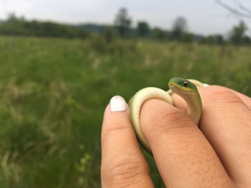
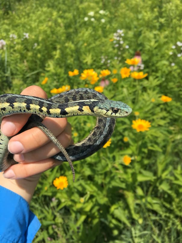
As mentioned above and before, snakes are beneficial to humans. How so, you ask? For one, snakes play a very important role in the ecosystem as both a predator and prey. As a predator, snakes keep rodent populations in check. In places that have removed snakes in the past, rat populations exploded and caused a lot of damage. Smaller species, such as green snakes, red-bellies, and young garter snakes also eat insects and can keep those populations in check as well. Snakes are a great method of pest control that is easy, harmless, and free. Please think twice about controlling pests with chemicals as that can damage the environment and harm other wildlife.
Along with eating rodents, snakes help control diseases that can increase when rodent populations get too large. Rodents carry several diseases that can be transmitted to humans and are hosts to ticks and fleas that can carry diseases like Lyme Disease, Ehrlichiosis, Rocky Mountain Spotted Fever, Plague, and Murine Typhus. Without snakes helping keep rodent numbers down, we could see increased prevalence of these diseases that are harmful to animals and humans alike.
As prey, snakes make a great meal for birds and larger mammals such as owls, hawks, fox, raccoon, and many other species. Believe it or not, some snakes, such as the Kingsnake and King Cobra, actually eat other snakes. These snakes are capable of eating venomous and non-venomous snakes. So not only do they help keep pest species populations from exploding, but they also serve as food for a number of other animals.
Lastly, snake venom is used in medicine. It contains hemotoxins that are used to treat heart attacks and blood disorders. The venom also contains neurotoxins that are used to treat Alzheimer’s, Parkinson’s Disease, strokes, and brain injuries. As the use of venom in medicine continues to grow, who knows how else venomous snakes can benefit humans.
If a snake bites your pet, try to move your pet away from the animal if it is safe to do so. Make sure to avoid getting bitten yourself. If possible, try to take a picture of the species to show your veterinarian. Bring your pet to an emergency veterinary center as soon as possible, do not wait. It’s best if you can call the veterinarian on the way so they are prepared to treat your pet as soon as you get there. Try to keep the bite below heart level, remain calm, and keep your pet calm as well.
If you suspect your pet was bitten by a snake but did not see it, here are some symptoms:
Swollen muzzle (will happen within 30 minutes of the bite)
Dual puncture wounds may be seen. They may or may not ooze red liquid.
Your pet may go into shock, of which symptoms include pale gums, mental dullness, increased breathing effort, slow breathing, or tremors.
How do I keep snakes out of my yard?
The two main resources that might attract a snake to your yard are food and shelter. Snakes eat rodents, fish, amphibians, and insects. By having snakes in your yard, they will keep populations of these animals low and help you avoid these animals. Snakes are great population control and are harmless, so having them in your yard is very beneficial. However, if you choose to make your yard unattractive to snakes, get rid of standing water, remove bird feeders, fill burrows around your house, and remove firewood, brush piles, tall grass, dense brush, and openings under sheds and concrete. Do NOT use chemical snake deterrents or traps, as these are either ineffective or pose a risk to other wildlife. The snake must also come into your yard for these products to be effective, which defeats the purpose of discouraging them from coming into your yard.
What should I do with an injured snake?
If you find an injured snake, it is likely from a predator attack, hit by a car, or from injury sustained during yard work. First, try to identify if the snake is venomous or non-venomous. If you are not sure, do not touch the snake. Also, before intervening, ensure the snake is sick or injured. Especially on sunny days, snakes find their way to gravel, pavement, or a warm flat space like a rock to bask. Usually, if you approach the healthy snake, it will move away into the grass. The snake is likely injured or sick if you see blood, scabbing, lesions on the skin, or it has a dull mentation, meaning it has lessened or no reaction to you or the environment. Once you have confirmed the snake needs assistance, you can use a stick or shovel to carefully move it out of harm’s way. If the snake is non-venomous, you can put it in a container with small air holes and a tight-fitting lid. If none are available, a pillowcase will also work. If the snake is venomous, do not touch it and call animal control. Keep the non-venomous snake in a warm and dark place until transport to a rehabilitation center. If the snake is stuck in a glue trap or netting, do not try to remove it. It is easy to further injure snakes in this position. Bring the whole trap or net to a licensed wildlife rehabilitator for removal as soon as possible.
How can I share my respect for snakes with others?
Through education and example. Tell others about why snakes are important, continue helping snakes in need, and let healthy ones go on their way when spotted in the environment! Knowing the benefit, they give to both humans and the environment may help convince people to at least leave a snake alone if they encounter one. Also, let people know that most snakes in our area are harmless and want to be as far away from humans as possible. Snakes only bite in self-defense and most venomous snake bites happen when someone is trying to handle or kill the animal. People don’t have to love snakes; we just ask that they respect them. In most cases, respect is just leaving them alone to continue on their way.
Every fall and spring, just like how thousands of students use the American highway system to return to home or school, hundreds of species of birds use a system of “flyways” to migrate according to the seasons. A flyway is an intricate pathway connecting all the regions birds use, including breeding, non-breeding, and staging areas. The American flyways can be roughly broken up into four distinct regions: the Atlantic Flyway, the Mississippi Flyway, the Central Flyway, and the Pacific Flyway. Flyways are complex and their borders are often hard to outline (parts of all the American flyways converge in Panama), but the birds that use them travel astonishing distances each year as part of their fascinating life cycles. Explore the flyways below and meet some of the charismatic species that use them!
The Atlantic Flyway
The Atlantic Flyway extends across the Eastern seaboard and contains iconic locations such as Chesapeake Bay, the Florida Everglades, and the Caribbean islands. The ecosystems in this flyway are incredibly diverse. From broadleaf forests and shrublands to coastal salt marshes, birds of all kinds depend on the natural resources across the region.
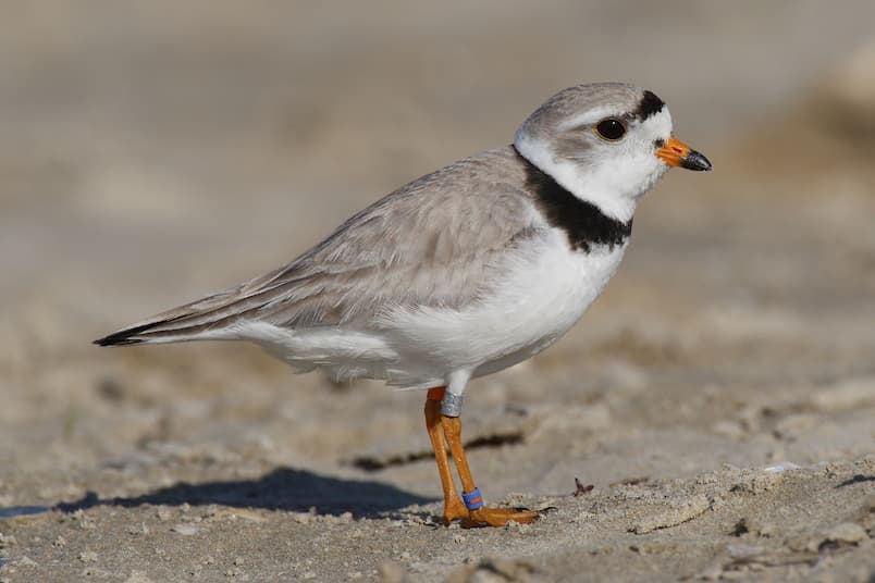
Adult piping plover, Image Retrieved via Wikimedia Commons, Creative Commons with Attribution
Piping plovers (Charadrius melodus) are regionally threatened or endangered and live on beaches, flats, and sandbars in coastal, Great Lakes, and Mississippi river waters. They are exclusively found in North America and are subject to the pressures of habitat loss and human disturbance. The birds are shy, preferring natural beaches without humans nearby. Conservation efforts have focused on protecting existing habitat and creating new habitat by dividing beaches to foster quieter, undisturbed habitat while still preserving recreational opportunities for people. As of 2020, their population numbers have been increasing. A pair actually nested on Montrose Beach in Chicago in 2019, the first time a nesting pair was seen in the area in half a century, and three birds were recently released by the Fish and Wildlife Service on that same beach this past July 2023.
The Mississippi Flyway
The Mississippi flyway follows the Mississippi river from its headwaters in Minnesota to its outlet into the Gulf of Mexico and across the gulf into South America. The migrating birds depend on the many prairies, wetlands, and forests found in the Mississippi river basin. The topography of this region is so flat that migrating birds do not need to adjust their height or direction to avoid geographical features like they do in the other flyways.
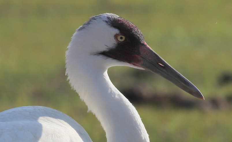
Adult whooping crane, Image Retrieved via Wikimedia Commons, Creative Commons with Attribution
The endangered whooping crane (Grus americana) utilizes the Mississippi flyway to travel from nesting grounds in Wisconsin, Iowa, Illinois, and other Midwest states south to wintering grounds in Texas, Louisiana, and Florida. The population reached a low of 15 individuals in 1942 and was estimated to be between 50 and 249 mature individuals in 2020. Migration patterns are a learned behavior in this species, posing unique challenges when trying to create new populations. To counteract this, researchers trained young cranes to follow an ultralight aircraft which was then flown across the United States from Ontario and Wisconsin down to wintering sites in Florida.
The Central Flyway
This flyway stretches from the Arctic to the Gulf of Mexico and connects the Rocky Mountains, the Southwest deserts, the great plains, and countless wetlands, grasslands, highlands, and desert ecosystems. The topography and varying climate causes migratory birds to take less direct routes to avoid the more inhospitable areas.
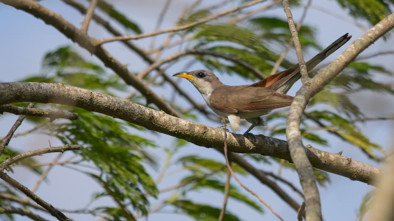
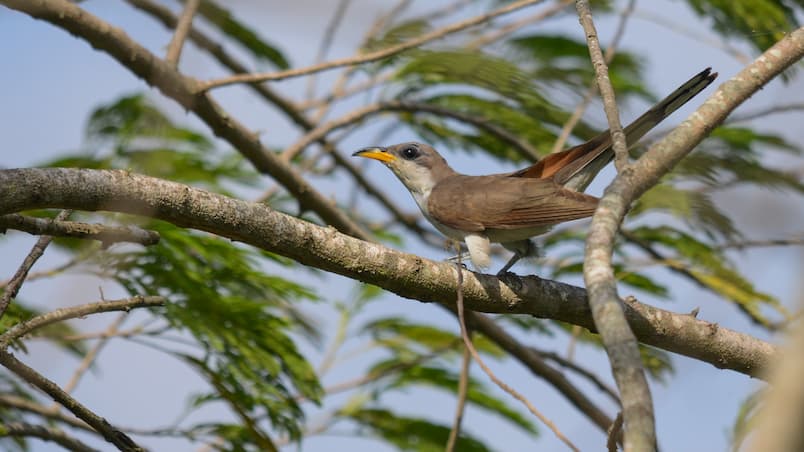
Adult yellow-billed cuckoo, Image Retrieved via Wikimedia Commons, Creative Commons with Attribution
When they hear the word “cuckoo” most people think of the European cuckoo, which is famously featured in cuckoo clocks made in the Black Forest region of Germany, but the cuckoo family of birds has members residing across the world, including North America. The yellow-billed cuckoo (Coccyzus americanus) can be found across much of the central and eastern parts of the United States, preferring riparian habitats (the edges of rivers, lakes, streams, and other bodies of water). Cuckoos are known for their strange and diverse calls, and the yellow-billed is no exception with at least six defined vocalizations including coos, knocks, kawlps, and croaks. Yellow-billed cuckoos are currently listed as a species of least concern but have been experiencing decline over the past few decades and are considered endangered in certain states. They have mostly disappeared from the westernmost states of the central flyway, as droughts and the building and use of dams have greatly degraded the available riparian habitat.
The Pacific Flyway
The Pacific Flyway includes Alaska, Washington, Oregon, California, and Hawaii and supports the migration of more than a billion birds across rainforests, coastal marshes, scrublands, deserts, mountains, and more. A larger network of state and federally protected lands in this region also provides more habitat for these species, lending to the abundance of wildlife seen throughout.
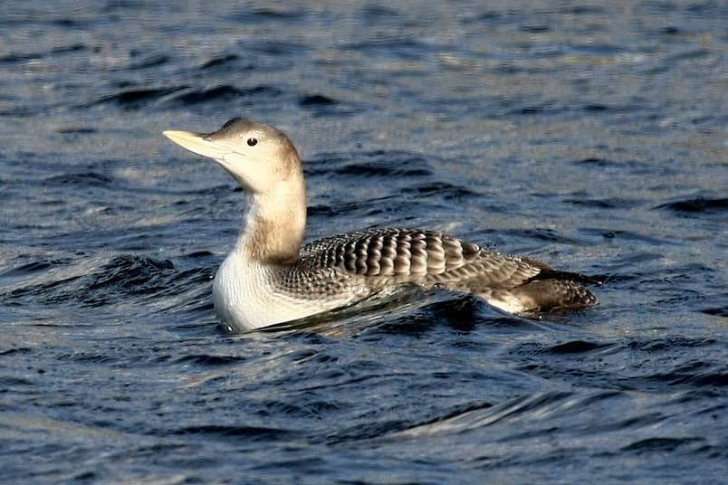
Juvenile yellow-billed loon – Image Retrieved via Wikimedia Commons, Creative Commons with Attribution
The yellow-billed loon (Gavia adamsii) is the largest member of the loon family which also includes the common loon, a rare but exciting visitor to the Wildlife Medical Clinic. Yellow-billed loons spend their summers in the high arctic tundra, fishing and breeding on the pristine lakes and rivers found throughout. They winter along the southern coast of Alaska and, rarely, along Washington, Oregon, and California. This species is extremely susceptible to pollution from oil and gas drilling and transport, as they depend on clean water and abundant sources of fish.
Loons have solid bones just like penguins and ostriches, but, unlike penguins and ostriches, loons are still capable of flight. The solid bones greatly decrease their buoyancy and aid in swimming, making them some of the best diving birds on the planet. These solid bones also mean that loons are heavy and awkward out of the water. Unlike other waterfowl such as ducks, loons are only capable of taking off from and landing in water, as they need a long wind-up directly into the wind to gain the lift they need. The increase in paved surfaces and the heat mirages they generate means that loons often mistake roadways as waterways and land on them. Without any water, they are then unable to take off again.
Just like all life on Earth, the flyways that migratory birds use are complex, beautiful, and not-entirely understood. In time we will unlock the secrets of the flyways, but it is important that we also protect the birds who depend on them. If you are still curious or would like to learn more about the flyways, you can find more information on them at www.Audubon.org/birds/flyways.
Summer is drawing to a close which means the Wildlife Medical Clinic is preparing for the return of our student volunteers! During the summer, the clinic is staffed by a team of 4 clinic interns, 2 student managers, undergraduate volunteers, and fourth year veterinary students. As classes begin in the fall, the clinic will welcome 115-140 student volunteers divided into 6 teams. Every student will be trained to provide care to the many species the clinic services.
Emydomyces testavorans is a recently discovered fungus associated with freshwater habitats that causes shell disease in a wide variety of turtles and terrapins (Woodburn et al 2019; Woodburn et al 2021). The fungus has been found in both wild and human cared for turtles and has been identified in individuals with active lesions and those with no evidence of disease.
E. testavorans was originally discovered in animals housed in captive care but recent studies found that it has the ability to infect and cause disease in wild populations as well (Lambert, MR, 2021).
Once established, lesions gradually eat away at the shell, progressing through the bone which also exposes turtles to infections. These lesions can have a wide variety of presentations from ulcerations of the shell, pitted depressions, discoloration and flaking of the shell, increased pliability of the shell, nodular masses within the shell or abdominal cavity, and cystic nodules.
The presence or absence of E. testavorans can be detected within shell tissues through polymerase chain reaction (PCR) run from swabs or scrapes of shell lesions and/or fresh or frozen tissue. Histopathology and necropsy can also be helpful in identifying whether an infection is present through stained shell tissues in the lab.
The recommended treatment for shell disease secondary to E. testavorans is still being investigated but nebulization of the anti-fungal terbinafine once daily for 28 days has been shown to be helpful, especially in addition to antifungal medication given over a set period of time and repeated in set intervals to help prevent reoccurrence. This is also known as pulse antifungal therapy. Other forms of treatment have included debridement of necrotic shell, packing lesions with terbinafine powder, and covering them with bone wax or cement.
Unfortunately, shell disease is a common cause of illness and death in turtles and the underlying cause for why some turtles will develop more severe disease remains unknown. Turtles can either be asymptomatic carriers or show active evidence of infection; however, those that show evidence of infection are more likely to succumb to severe, chronic disease of the shell and a large number of those severely infected turtles will die.
Identifying the fungus is an important first step which allows us to better understand how the fungus spreads which can help us to develop health management strategies for impacted turtles, and ways to combat potential emerging disease. Having this knowledge provides us with not only the means to quickly diagnose patients and implement possible treatment options, but is also essential in assisting to develop proper handling and biosecurity measures as well.
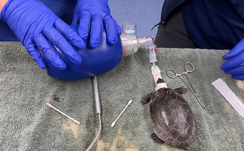
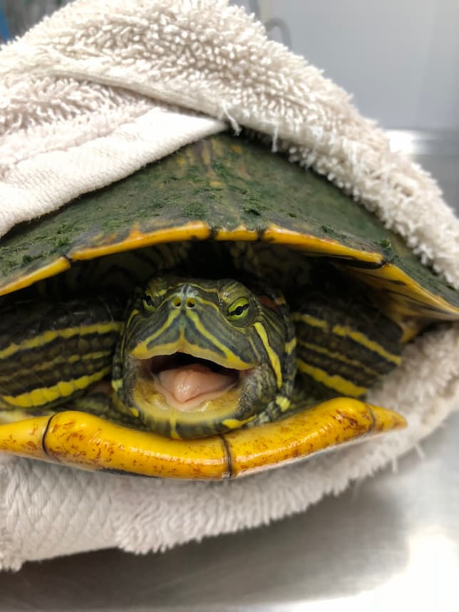
*Turtle images featured in this article are for visual design. They do not have the disease mentioned.
Written by Tori McCarthy, Affiliate Clinical Year DVM Student
The Wildlife Medical Clinic at the University of Illinois College of Veterinary Medicine embraces a threefold mission: to provide veterinary care to sick or injured wild animals; to educate veterinary students and wildlife professionals about wildlife medicine; to promote conservation initiatives and ecosystem health through collaborative research, dissemination of scientific information, and public education programs.
Read our annual report to learn more about how we have carried out our mission throughout the year!