Feline Thorax Example 1
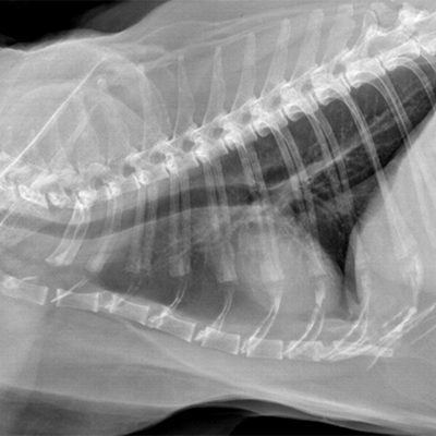
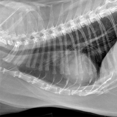
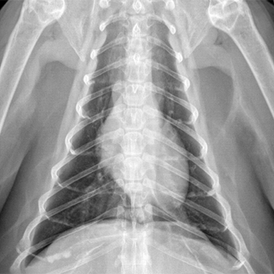
The following radiographs are the left lateral, right lateral and ventrodorsal views of the thorax of a seventeen-year-old mixed breed cat. Degenerative changes are present within the costal cartilages and elbows in addition to possible joint capsule mineralization present in both elbows.
Click images below - interactive images will open in a new window