Equine Skull Example 4
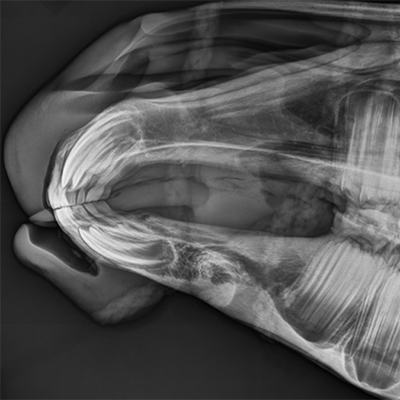
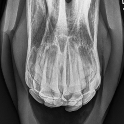
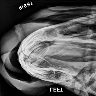
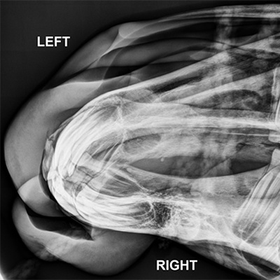
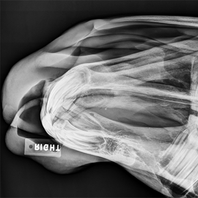
The following radiographs are the left lateral and dorsoventral views as well as two dorsoright-ventroleft oblique’s, one dorsoleft-ventroright oblique, & an intraoral maxillary radiograph of the skull of a two-year-old thoroughbred filly. There exists a slight incisor tooth malalignment resulting in ulceration over upper left first incisor.
Click images below - interactive images will open in a new window