Canine Skull Example 1
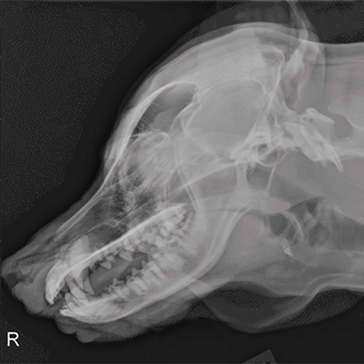
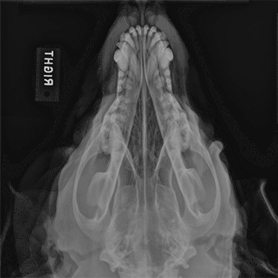
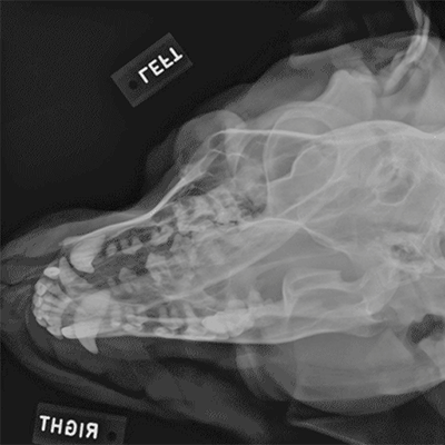
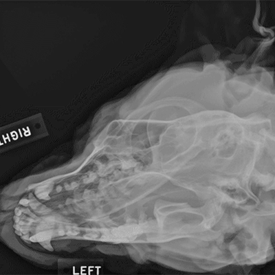
The following radiographs are the right lateral view of the skull and neck as well as dorsoventral, dorsoventral right-left oblique and dorsoventral left-right oblique views of the skull of a ten-year-old Labrador Retriever. On the dorsoventral view there is increased soft tissue present lateral to the right zygomatic arch and superimposed over the external ear canal and pinna.
Click images below - interactive images will open in a new window