Canine Hindlimb Femur Example 1
The following radiographs are the mediolateral and craniocaudal views of the right femur of a one-year-old Standard Poodle.
Click images below - interactive images will open in a new window
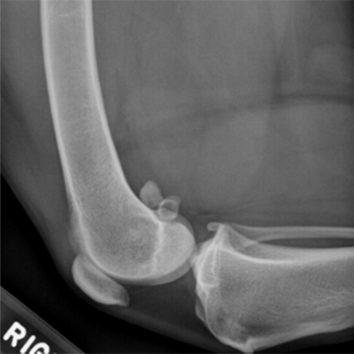
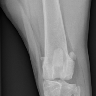
The following radiographs are the mediolateral and craniocaudal views of the right femur of a one-year-old Standard Poodle.