Canine Hindlimb Femur Example 5
The following radiographs are the right lateral and ventrodorsal views of the pelvis and femurs of an eight-year-old Mixed Breed dog.
Click images below - interactive images will open in a new window
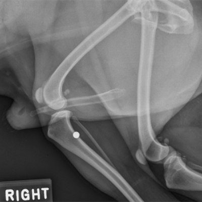
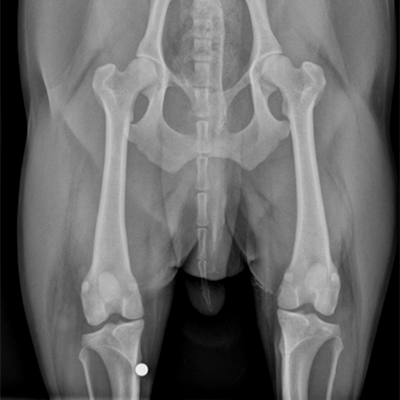
The following radiographs are the right lateral and ventrodorsal views of the pelvis and femurs of an eight-year-old Mixed Breed dog.