Canine Abdomen Example 4
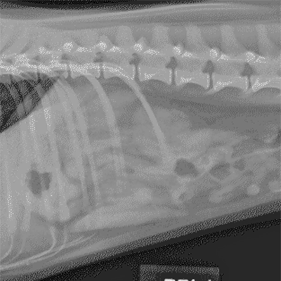
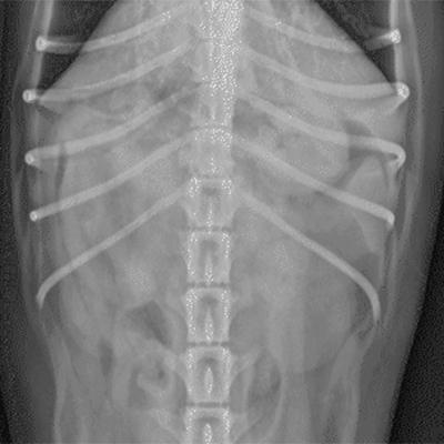
The following radiographs are the left lateral and ventrodorsal views of the abdomen of a two-year-old Mixed Breed Dog. On both the lateral and ventrodorsal view, a large calculus is present in the urinary bladder; it measures approximately 2.5 by 2 cm. There also exist two metallic sutures present in the ventral floor of the abdomen.
Click images below - interactive images will open in a new window