
Recently in the field, we encountered numerous turtle shells left behind from previous mortality events. While these discoveries can initially feel discouraging, they also represent an important opportunity for wildlife disease surveillance. Even when soft tissues are long gone and all that remains is shell and bone, there is often still valuable information preserved that can help us better understand mortality events and disease dynamics in wild turtle populations.
One of the pathogens we are particularly interested in investigating is Ranavirus, a highly significant pathogen of reptiles, amphibians, and fish that has been associated with mortality events in turtles, including eastern box turtles. Ranavirus causes a systemic infection, meaning it spreads throughout the body rather than remaining isolated to a single tissue or organ. Because of this systemic distribution, traces of viral DNA may still be present in tissues like bone marrow even after substantial decomposition has occurred.
This is where shell sampling becomes incredibly useful.
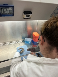
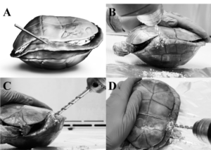
Drilling shells at WEL to harvest bone marrow
Rather than relying on fresh tissues (which are often unavailable in field mortality investigations) we can collect samples directly from the shell itself. In the lab, a drill is used to carefully access and harvest remaining bone marrow material preserved within the skeletal structures. Although the animal may have decomposed long ago, these internal tissues can still contain DNA from pathogens present at the time of death.
After collection, the bone marrow samples undergo DNA extraction, where genetic material is isolated from the tissue. The extracted DNA can then be analyzed using PCR (polymerase chain reaction) techniques. PCR acts as a molecular detection tool by amplifying tiny amounts of target DNA sequences so they become detectable. For Ranavirus surveillance, PCR assays target viral genetic regions to determine whether viral DNA is present in the sample. Quantitative PCR (qPCR) methods can even estimate viral loads when enough material is available.
This approach is especially important because wildlife mortality events are often difficult to investigate. Animals may not be discovered immediately, carcasses decompose rapidly, scavengers remove tissues, and sometimes the only evidence left behind is a shell. Historically, many of these cases would have been impossible to evaluate.
https://pubmed.ncbi.nlm.nih.gov/29297832/
Research by Claire E. Butkus, formerly a WEL student and now a wildlife student with California Department of Fish and Wildlife, helped demonstrate the value of this technique. Her work showed that bone marrow harvested from skeletonized eastern box turtle shells could successfully be used to detect Frog Virus 3 (FV3)-like ranavirus using quantitative PCR. In their study of 87 shells, ranavirus DNA was detected in bone marrow samples from multiple turtles, supporting shell-derived bone marrow as a practical tool for postmortem surveillance of systemically distributed pathogens.
Fieldwork is not always live captures, nesting surveys, or healthy animals. Sometimes it means finding the remnants of past events and asking what they can still teach us. Through methods like shell drilling, bone marrow harvesting, DNA extraction, and PCR testing, even a shell can continue contributing to conservation, disease surveillance, and our understanding of wildlife health long after the animal is gone.