
What am I doing here? What is the meaning of all of this? Ok, don’t worry, this is NOT an existential crisis. I thought I would take a minute this week to show all of you the process I go through when processing box turtles! Each turtle has a different personality and varying levels of stubbornness during sampling, but the methods are generally the same.
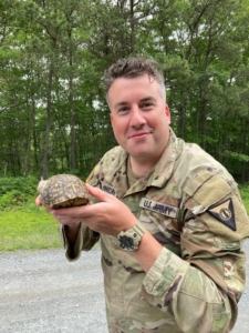
Corporal Johnson holding a box turtle he found on the road!
Step 1: Find the turtle! – This step is the most challenging and time-consuming. Most of the box turtles I’m sampling have transmitters, so we trek through the woods to find them with radiotelemetry. Sometimes we get lucky and happen to stumble across new ones or get calls from soldiers on base that have found box turtles on their own! These are definitely the most exciting since I get to process them, and the field techs get to place a new transmitter to add the turtle to our squad. At the location where the turtle is found, a GPS point is taken and some initial data such as air temperature, humidity, substrate temperature, etc. is recorded. All equipment is also appropriately disinfected between turtles to prevent cross-contamination.
Step 2: Physical Exam – This is where the real fun begins! Depending on the personality of the turtle, this step can be brief or take the majority of the time. Some turtles are super social and like to see what’s going on outside their shell. These are the BEST turtles to work with since I can easily get a good look at their entire body for a quick and thorough exam. Other turtles are shy and like to stay boxed into their shells. I still love these turtles, but they do make my job a little harder. For these stubborn turts, I use a probe to open their shell, and then place a syringe case inside to keep them open for the duration of sampling. If you ever want to be truly humbled by how weak you really are, try and get a box turtle open… These guys are WAY stronger than they appear and have me wishing I spent more time in the gym before starting this fieldwork.
Step 3: Draw blood – Before we take blood, we need to do a little bit of math. No more than 0.8% of a turtle’s body weight is taken in blood, so this calculation is always done prior to venipuncture. We draw from the subcarapacial sinus which sits under the front of the shell, right behind their head. This location is fairly reliable for drawing blood, except for the occasional lymph contamination from the network of lymph vessels that sit in the same area.
Step 4: Put blood into tubes – This step needs to be done quickly in order to prevent the blood from clotting in the syringe. I’ve found that quietly saying “please don’t clot, please don’t clot, please don’t clot” to myself as I put the blood into tubes usually does the trick. We use tubes lined with lithium heparin to prevent coagulation, and another one with a solution to preserve RNA.
Step 5: Swabs – We take 4 types of swabs: oral, oral-cloacal, cloacal, and shell. For our feisty turtles, the oral swab is no problem, since they usually take the opportunity to chomp down on the swab once it gets close to their mouth. The cloacal swab is a little more invasive, but still fast and easy. Finally, the shell swab is a nice back massage over the entire carapace & plastron.
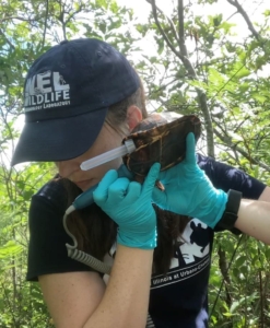
Taking a heart rate with ultrasound
Step 6: Put swabs into tubes – This year we have an array of brightly colored Eppendorf tubes, so each swab has a specific color. The swabs are broken so they fit in each tube, and then placed in order for each individual turtle. After sampling 100+ turtles, I have an entire rainbow of tubes all waiting for DNA/RNA extraction once I get back to our lab in Urbana.
Side 7: Take shell measurements – Using exceptionally large calipers (that like to get caught on every low-hanging branch as I hike), measurements of the carapace and plastron are taken. These numbers, in combination with the turtle mass, are used to calculate a body mass index.
Step 8: Take heart rate with ultrasound – A portable vascular ultrasound is used to detect flow of blood in vessels and in the heart. For larger turtles, the probe can be placed on the skin between the turtle’s neck and forelimb. For smaller turtles or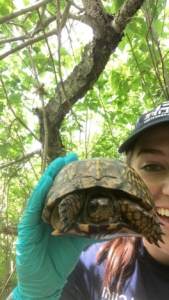 juveniles, the probe is placed on the middle of the plastron. Ultrasound gel is used to help conduct sound, and the characteristic “whooshing” noise is counted to get a heartrate in beats per minute.
Final (optional, but most exciting) Step: The Turtle Selfie – Because duh. These turtles are infinitely photogenic and adorable.
There you have it, folks! Tune in next week to find out what happens with the samples once we get back to the lab 😊
Total Turtle Count = 108