Toxicology and Environmental Health Training Program
The University of Illinois’ Research Training Program in Toxicology and Environmental Health is a part of the larger, campus-wide Interdisciplinary Environmental Toxicology Program (IETP), which provides toxicology training to students and postdoctoral fellows trained in basic sciences such as endocrinology and reproductive biology. Our program is ideal for students who are interested in applying their basic knowledge in four areas of toxicological research: reproductive/endocrine toxicology, neurotoxicology, nutritional toxicology, and nanotoxicology. The Research Training Program is a T32 NIEHS Environmental Toxicology Training Program.
Faculty
TRAINING PROGRAM FACULTY PRECEPTORS


Interdisciplinary Environmental Toxicology Program (IETP)
The IETP Program at the University of Illinois provides individuals from many disciplines with a basic understanding of the complex interactions between chemicals and living organisms. Created in 1984, it combines the highest standards of scholarship with a flexible program that enables graduate students from departments and colleges across the campus to specialize in this exciting field.
What is IETP?
Students in biology, chemistry, engineering, entomology, microbiology, neurosciences, nutrition, physiology, veterinary medical sciences and other graduate programs at the University of Illinois may specialize in environmental toxicology. The program is rigorous, demanding that students fulfill the degree requirements of their own departments as well as the additional requirements of the program. The program includes course work in toxicology and an interdisciplinary seminar series that features guest speakers from academia, industry and government. The student’s independent thesis or dissertation research must address a toxicological problem.
For admittance into the program, students apply to a participating department and contact one of the program’s faculty about serving as a research advisor. Graduate students who believe their research will benefit from being associated with the program should discuss this possibility with their advisors, regardless of whether the advisor or department is associated with the program.
Faculty
The following faculty, listed alphabetically, participate in the Interdisciplinary Environmental Toxicology program by teaching courses that meet program requirements, engaging in research on toxicology-related subjects, advising graduate students, and participating in a variety of program activities on a voluntary basis. Names and photos are linked to corresponding webpages.


Seminar Series
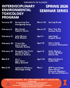
Interdisciplinary Environmental Toxicology Program Seminar Series
Spring 2026 IETP Seminar Series Schedule
Current Students


Stipends
Stipends for predoctoral and postdoctoral trainees are for two years (one-year, renewable pending satisfactory performance).
Predoctoral Trainees
- The Stipend is $28,788 per year
- Tuition and some fees are covered
- Travel allowance ($500) to a scientific meeting
Postdoctoral Trainees
- Travel allowance ($600) to a scientific meeting
- Subject to funding availability and current stipend scales as dictated by NIH.
- Stipends depend upon years of experience:
- 0 — $62,232
- 1 — $62,652
- 2 — $63,120
- 3 — $65,640
- 4 — $67,824
- 5 — $70,344
- 6 — $72,960
- 7 or more — $75,564
Coursework
Core Course requirements for the Interdisciplinary Environmental Toxicology Program are flexible to accommodate the varied career goals of our students.
Three courses are required:
Interdisciplinary Toxicology Seminar
CB 596 (1 hr. every semester): Topics will vary each semester. Seminars are presented by faculty, visiting lecturers, and students based upon their study, research, and/or professional activities in the selected topic area. Same as ENVS 596, and PATH 596. See PATH 596. Offered Fall and Spring ever year.
Systems Toxicology
CB 554 (3 hrs.): This course is designed to provide an overview of the effects of chemicals and their mechanisms of action in a variety of organ systems. Topics include toxicology of the nervous, developmental, reproductive, thyroid, renal, hepatic, immune, pulmonary, and gastrointestinal system. Offered Spring of odd years.
Ethics in Toxicology
CB 552 (2 hrs.): Emphasis on ethical issues in the practice of toxicological research: collaboration, authorship and plagiarism, professional responsibility to subjects (both human and animal), whistle-blowing, codes of ethics, legal obligations. Featuring case studies. Offered Fall of even years.
Strongly suggested courses include the course selections below:
Biochemistry/Chemistry
(6 hours or 2 units) Specific courses will depend on requirements of advisor’s home department.
Biostatistics
(3 hours) Specific courses will depend on requirements of advisor’s home department.
Basic Toxicology
CB449, ENVS 480 (3 hours): Emphasis on physiology and biochemistry of intoxication; discusses the types of cellular response to toxic compounds and the role of species variation in toxic responses.
OTHER AVAILABLE COURSES IN TOXICOLOGY:
CB 551 – Ecotoxicology in the Northern Hemisphere, Offered each Fall semester
CB/ENVS 514 – Neurotoxicology, Offered Spring of odd years
CB/ENVS 516 – Reproductive and Developmental Toxicology, Offered Spring of even years
CHLH 572 – Principles of Epidemiology in Public Health
CHLH 578 – Applied Epidemiology
CHLH 576 – Analytic Epidemiology
ENVS 469 – Environmental Health, Offered each Spring semester
ENVS 491 – Sustainability Experience
FSHN 575 – Issues in Food Safety
MCB 529 – Scientific Writing
NUTR 550 – Grantsmanship and Ethics
PATH548 – Toxicologic Pathology
You may learn more about each course through the University Course Catalog.
Contact.
Let us know if you have any questions about our programs
Jodi Flaws, Director
College of Veterinary Medicine
Department of Comparative Biosciences
3223 Comparative Biosciences
Urbana, Illinois 61802
MC-002
Phone: 217-333-7933
Email: jflaws@illinois.edu
Lori Raetzman, Associate Director
School of Molecular & Cellular Biology
College of Liberal Arts & Sciences
University of Illinois at Urbana-Champaign
407 S. Goodwin Ave.
Urbana, Illinois 61801
Phone: 217-244-6233
Email: raetzman@illinois.edu
Synthia Lane, Program Coordinator
Toxicology Training
College of Veterinary Medicine
2001 S. Lincoln Ave.
Urbana, IL 61802
217-300-6465
Email: sjlane@illinois.edu