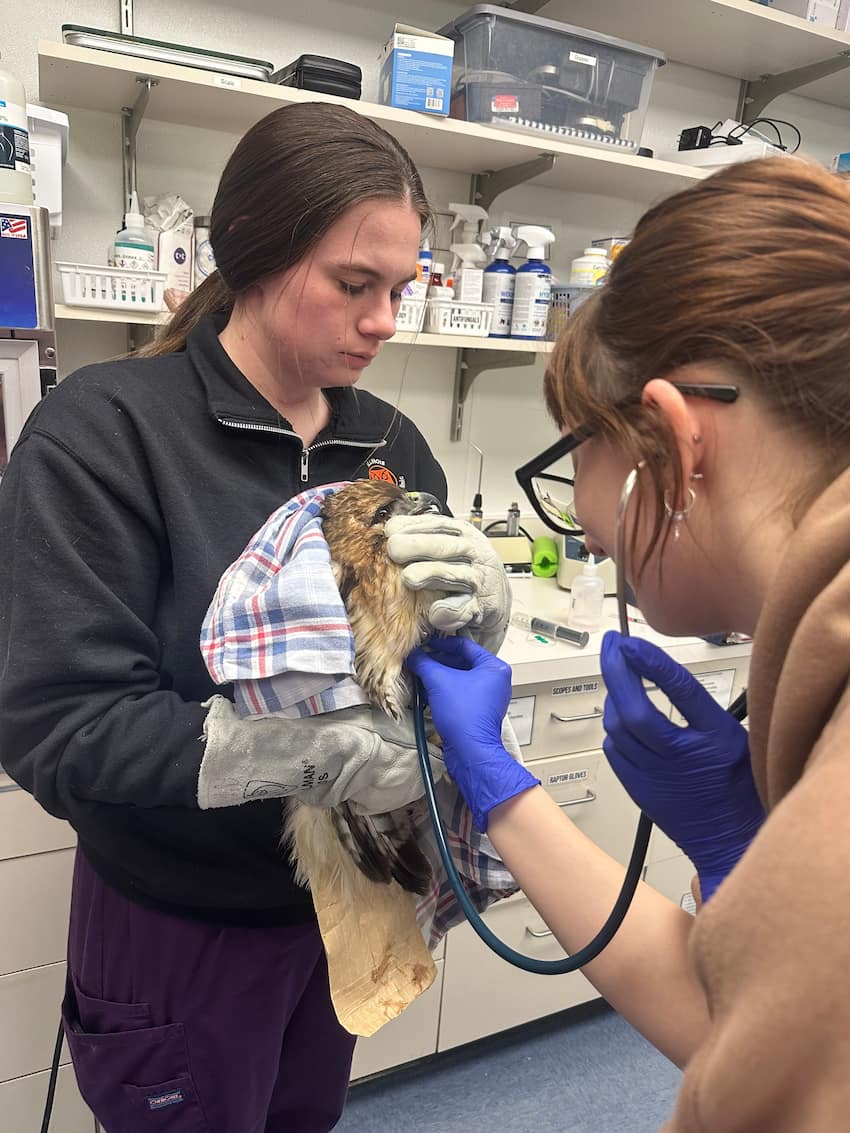
Observation and Diagnostic Tools
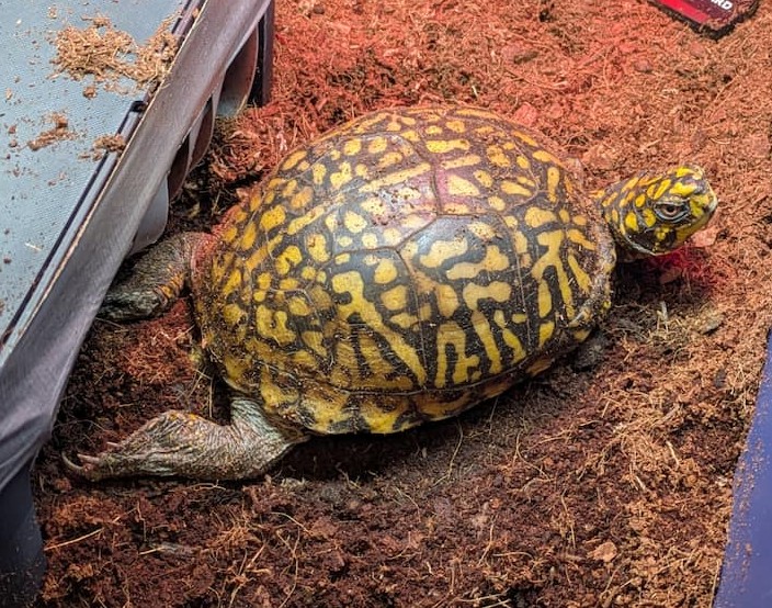
When a good Samaritan brings in a Great horned owl that cannot fly or a lethargic Eastern box turtle with no apparent physical injuries to the Wildlife Medical Clinic, how do we determine what is going on with these patients? That’s where the problem-solving aspect of our work comes into play. In all fields of veterinary medicine, our patients cannot speak for themselves, and we must rely on clinical signs as well as the owner’s perspective of their pet’s behavior. However, wildlife patients are a little different in that they do not have owners who can speak for them. There are many different diagnostic tools we can utilize to aid in our understanding of what is going on with our patients, such as bloodwork and radiographs.
Bloodwork
In our bird and reptile species, bloodwork can be challenging to both perform and interpret due to a lack of reference intervals for every species and differences in cell morphology. Mammalian species have red blood cells that lack a nucleus. Birds and reptiles are special in that their red blood cells have a nucleus. This means that automated machines cannot accurately differentiate and count red blood cells and white blood cells. Anytime a blood sample is collected from a patient in the clinic, we want to manually determine a packed cell volume (PCV) and a total protein value (TP). We will also make a blood smear on a slide to later be evaluated under a microscope. In order to get the PCV and TP, we must first centrifuge the sample in a micro-hematocrit capillary tube which will separate the sample into 3 distinct layers. Red blood cells are at the bottom, white blood cells and platelets in the middle, and plasma at the top. We use a special card to aid in reading the PCV, or the percentage of red blood cells that are in the sample. Total protein is then measured by placing a drop of the plasma onto a tool called a refractometer. PCV and TP values can give us a clue on the hydration status of our patient as well as if they are anemic at that time. The blood smear mentioned before is used to evaluate cell presence and morphology. The presence of specific white blood cells can give us clues on if the patient has inflammation or even if there are microbial organisms within the cells. However, a challenge we often face in wildlife medicine is determining what values are normal as there often are no reference intervals available for many of the species we work with. Sometimes, we must make inferences based on what we already know about closely related species.
Radiographs
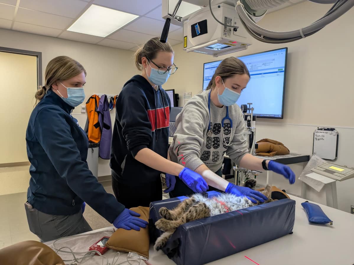
Another helpful tool we utilize often in all fields of veterinary medicine are radiographs. Any patients presenting with suspected orthopedic injuries will have images taken to be further assessed. We also take radiographs of birds who present with the inability to fly but have no obvious injuries to see if there are any fractures or dislocated joints. Radiographs can be helpful to determine the location and extent of these injuries so it can be decided whether or not the injury can be fixed. Imaging is also helpful for evaluating abnormalities in internal structures and can sometimes highlight the presence of metals that may have been ingested by the animal.
Conclusion
These diagnostic tests allow us to get one step closer to fixing our patients so they can be released back into the wild. Bloodwork and imaging are only two of the many tools available to us at the Wildlife Medical Clinic. If we suspect a specific disease, we can often run a specific test to either confirm our suspicions or rule that out. A closer look at what is happening inside of our patients, whether that is in their blood, skeletal system, or internal structures, can help guide us when creating treatment plans and determining a prognosis for our patients.
Written By: Grace M., Class of 2028