Summer is the busiest season in the Wildlife Medical Clinic! To ensure we provide quality care and support to our patients, our summer interns have been putting their whole heart into caring for our wildlife patients and supporting our faculty, house officers, and 4th-year vet students. Each year, we hire 3-4 veterinary medicine students, who have been involved in the clinic for at least a year, as summer interns. Their main responsibilities are to provide near-peer mentoring to our 4th-year students on case management, medical treatment, diagnostic choice, and all things wildlife. Below are some of the case highlights from each of our summer interns this year and their reflections on the season!
Rachel Eggleston, Class of 2027- Case Highlight:
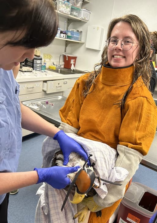
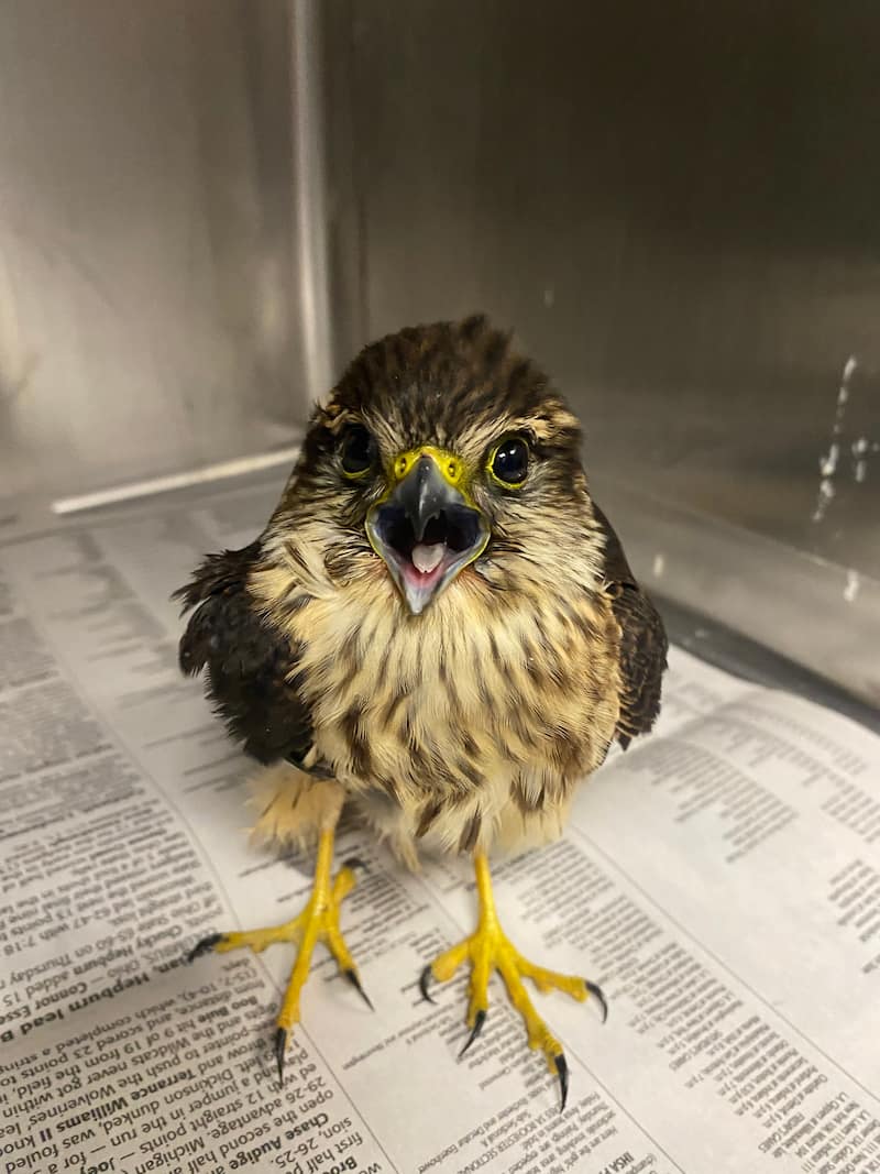
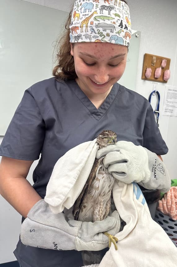
At the beginning of August, the Wildlife Medical Clinic received a female American Kestrel. She came to us after being found near the side of the road and was not acting normally. I do not have a lot of experience with American Kestrels in the clinic and I really enjoyed getting to be involved during the triage exam for this patient. During her physical exam, there were not any obvious injuries or abnormalities, but we did notice some increased laxity in her right wing. We also looked into her eyes with an ophthalmoscope and were able to see evidence of minor hemorrhage or blood in the right eye. While she has been in the clinic, we have been providing pain medications, anti-inflammatories, and additional supportive care as needed. I look forward to working with her more and seeing her progression as she continues to heal!
Rachel’s Thoughts
My name is Rachel Eggleston, and I was chosen to be one of the DVM student interns for this summer! I will be going into my second year of veterinary school this fall. Some of my favorite memories from this internship have been getting to work with different raptor species. One of my favorite memories that I will remember for a long time is getting to scrub in and suture close on a surgery for a Great Horned Owl! I have always been a fan of birds and thoroughly enjoyed helping treat a Bald Eagle we had in the clinic for a while before it was transferred to a different wildlife center. I love so many things about being able to work in the WMC, but I think what I value the most from my time this summer is the long-lasting connections I have been able to make. I have greatly enjoyed getting to work alongside clinical year students, faculty, residents, and the WMC managers. This summer program has allowed me to grow and strengthen my skills as a future veterinarian, student, and colleague, and I am so grateful to have had this opportunity!
Roxanne Lisowsky, Class of 2027- Case Highlight:
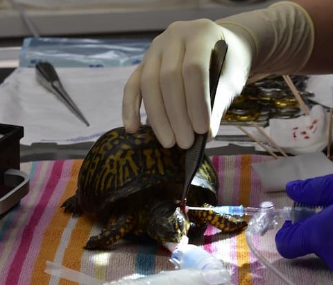
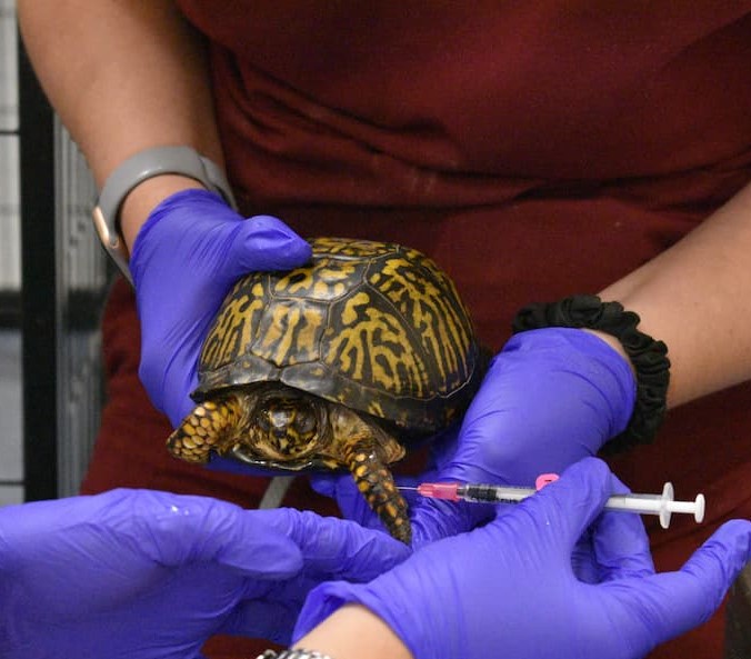
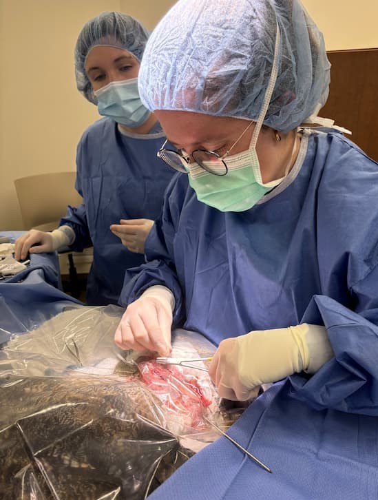
In early July, we had an adult female eastern box turtle come into the clinic for swelling on the sides of her face and not moving around much. Upon our initial exam we found that the patient had aural (ear) abscesses on both sides. The turtle also had some crusting and discharge in both eyes along with nasal discharge. We were able to drain the larger abscesses and started her on antibiotics to help clear the rest of both abscesses. We also gave pain medication and anti-inflammatory medication to help with her healing. During her stay we also found she had corneal ulcers that we treated with eye medication, and they resolved. We had to drain the abscess one more time during her recovery. We also tested for mycoplasma, and it came back negative while the culture of the abscesses came back sensitive to the antibiotic we used. She continued healing and recovering well in the clinic. Turtles heal very slowly, and it takes a lot of patience and dedication to help them along the way. We made sure she finished her course of antibiotics and then she was given one last exam to confirm her clean bill of health before being released on August 9. We hope she has a long and healthy life!
Roxanne’s Thoughts
My favorite part about this summer has been working with the undergrads and 4th-year students. I love being able to show them new things about wildlife patients, teaching new skills, and seeing the lightbulb moments. I grew more confident in my daily clinical abilities and feel prepared and excited to be a team leader for the next school year. I also enjoyed seeing patients we don’t always get during the school year.
Caroline McCormick, Class of 2027- Case Highlight:
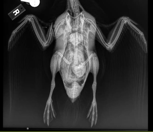
An adult female mallard presented to the wildlife medical clinic on May 29, 2024, with an acute mid-diaphyseal long obliqued simple right humeral fracture, opened on ventral aspect with ~1-2 cm of exposed bone with each fragment. The injury appeared very acute (< 6 hours old), and not grossly contaminated. There was relatively minimal muscle/soft tissue damage.
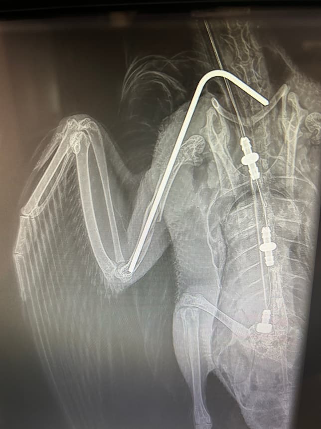
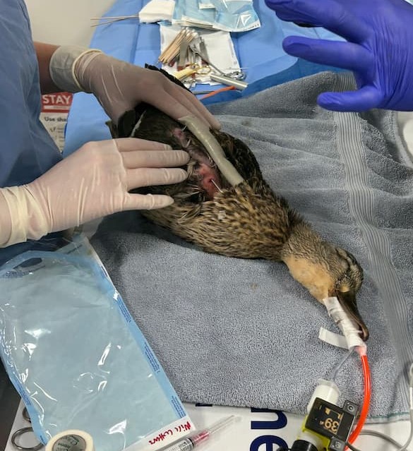
Immediately upon intake, radiographs were performed to confirm the injury, and the animal was prepped and taken to surgery. An inter-medullary pin was retrograded into the right proximal humerus via existing the ventral wound. The distal fragment was aligned and the IM pin advanced until it was well seated in the distal fragment. The open fracture was closed with a continuous horizontal mattress suture. The IM pin was bent using a chuck and vice grip and then blunted. A body wrap was placed to stabilize the wing for healing. The mallard was then started on pain medications and antibiotics.
Physical therapy was performed three times per week for several weeks to prevent loss of range of motion in the wing. The pin was slowly destabilized or removed as the wound healed. Repeat radiographs were performed to monitor healing. Eventually the pin was removed completely. Once the mallard was healed, she was transferred to another facility for continued rehabilitation before release.
Caroline’s Thoughts
The summer program has been wonderful. I have enjoyed working with so many wonderful people and animals. I have learned a lot and been able to practice many skills.
Sofia Murillo Logan, Class of 2026- Case Highlight:
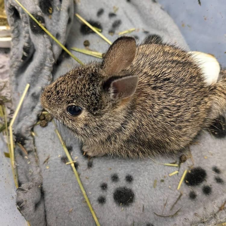
One of the most inspiring cases from this summer is from a small bunny that came into the Clinic. An unidentified juvenile eastern cottontail presented to the Wildlife Medical Clinic on June 14, with a history of a broken left leg. On the intake exam we found a fracture of the right tibia, but other than that, this bunny was pretty healthy. Now when animals come into the clinic there are a list of factors we always need to consider for the health and safety of the animals and the volunteers. “How will this animal respond to captivity while in the clinic, and what is this animal’s prognosis.” Prognosis meaning based on everything going on with the animal and the road they face ahead, how likely are they to survive to then be released.
When speaking to Dr. Lewis about this case and evaluating the prognosis of this patient we decided to keep this patient for further evaluation. Due to their good body condition and the stability of the fracture we created a plan for this patient. Body condition can be a huge aspect to the prognosis of a patient. This comes down to, how stable is this animal, and if it is not stable, how long will it need to become stable. Without the patient being in good body condition and having appropriate neurological signs, it can be hard to treat severe injuries successfully.
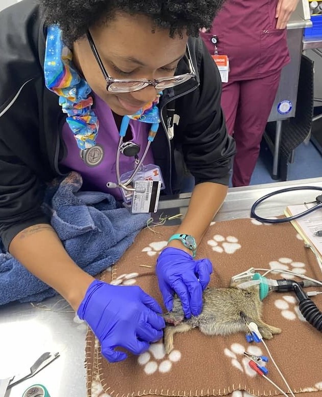
Throughout the patient’s stay the bunny was doing very well. We stabilized the fracture site with an Ehmer sling, the same process as when you need to put your arm in a sling when you shouldn’t use it, for healing purposes. We kept the bunny on some pain medications to keep them comfortable while in our care. We took radiographs (x-rays) on the patient to visualize the fractured leg. After confirming the stability of the fracture, the doctors were able to fix the leg surgically by placing an intramedullary pin. This is a pin just a little smaller than the width of a bone that is used to keep the bones in place while the patient is healing. After surgery our patient was still doing well and gaining weight, which again eating is a positive prognostic indicator. After about a week we checked to see the healing progress of the patient by taking another radiograph. Since there was evidence of a bony callus starting to form, we removed the sling and allowed the patient to freely move around the cage to encourage movement of the stifle (knee) and hock (ankle). After another week another radiograph was taken to see if it was time to take the pin out of the patient’s leg. Due to the stability and healing of the leg we did decide to take the pin out. The pin was removed a little over 2 weeks after being placed. We were able to remove this pin this early because bunnies can heal very quickly, especially young ones. We also didn’t want the pin to limit this growing bunny’s leg, so we were very happy to remove it so quickly. The bunny was able to be released a few days after pin removal.
Overall, the patient did very well and gained over 100g during their time with us in the clinic. Something we could have done differently in this case was to keep this patient a little bit longer to get more strength built in the injured leg but ultimately it was doing well, and we felt comfortable with releasing when we did. We were very happy with the result of this case and hope this eastern cottontail is out in the wild eating all the greens and growing to adulthood!
Sofia’s Thoughts
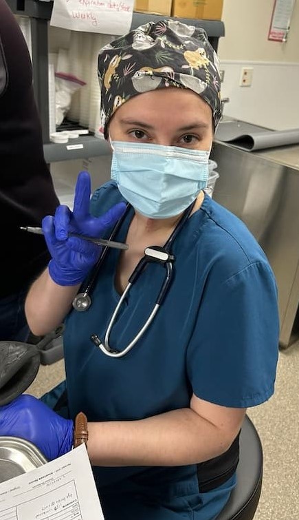
My name is Sofia Murillo Logan and I am one of the Wildlife Medical Clinic Summer Interns. I applied to this internship because I wanted to spend more of my time immersed in wildlife. Volunteering for the clinic in the school year can be difficult due to the rigors of veterinary school. My experience thus far has been nothing short of inspiring. I get to learn daily from our amazing WMC Director, Dr. Stephany Lewis, as well as our house officers that are working towards specializations. Being at the clinic has increased my confidence and knowledge not just in wildlife species, but also in medicine and clinical reasoning. I have greatly enjoyed working with our fantastic and knowledgeable managers Tyson Jenkins and Riley Dunwoody. They both are incredible leaders and are passionate about doing what’s best for all the creatures in our care, as well as for the students in the clinic. For any students at the University of Illinois interested in gaining more hands-on experience I highly recommend getting involved in the Wildlife Medical Clinic.
Written By: YiYing and Sofia, class of 2026, and Rachel, Roxanne, and Caroline, Class of 2027.