Tony Bieser, VM20, Team Leader
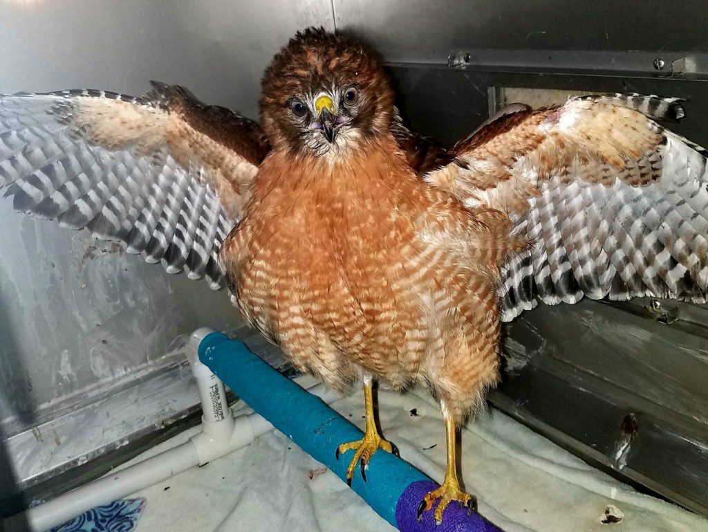
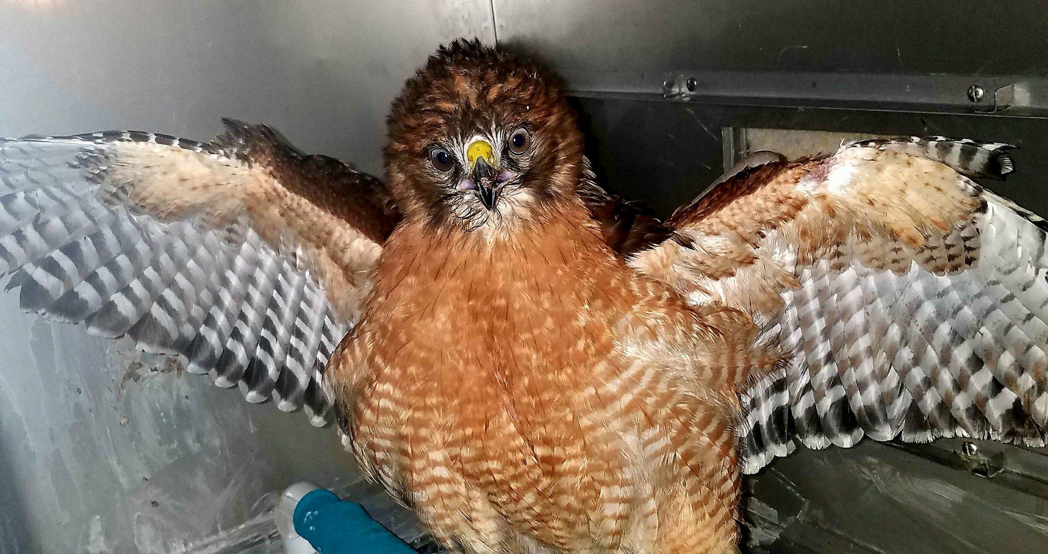
Red-shouldered hawk (Buteo lineatus)
This hawk presented to the Wildlife Medical Clinic after being found down near a public road. The finders cared for the patient for an unknown amount of time before they eventually brought them to the WMC for care. On presentation, the RSHA had an obvious left-wing deformity and was unable to fully extend their elbow and carpal joint. Because of this abnormality, radiographs were performed to determine etiology.
Pre-op Radiographs: The image to the right depicts a healing, mid-diaphyseal fracture
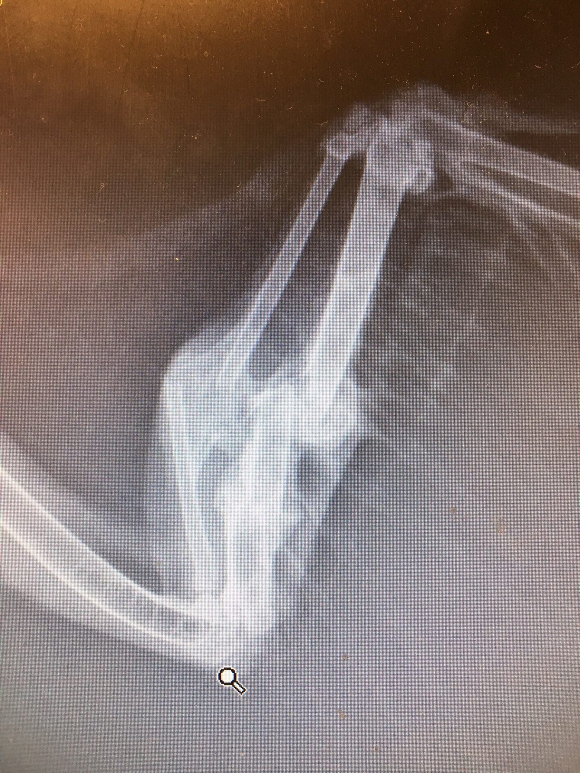
of the patient’s left radius and ulna. Upon closer inspection, there is loss of detail in the elbow and carpal joints as well. Fibrous tissue appears to be forming between the radius and ulna, and a callus is present between the fracture sites of each bone.
*Note: The radius and ulna must be able to move independently of each other for the range of motion needed for flight. When both bones are broken, they are at risk of healing together and bridging (like in this patient). This new bone formation prevents the radius and ulna from sliding past each other, and prevents the bird from flying.*
Due to the malalignment of the fracture, surgery was performed to allow for more appropriate healing.
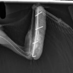
Surgery: This patient underwent surgical correction using intramedullary (IM) pin placement in both the radius and ulna. The fibrous tissue formed between the bones was broken down to ensure that both bones were free from one another (the bones must be able to glide independently of each other for flight). An IM pin was placed in each bone and three additional pins were placed on the outside of the patient’s wing to serve as part of an external fixator. The external fixator helps prevent the IM pins from moving, which will help the patient’s fractures heal in the proper position. Patient was placed in a body and wing wrap for three days until post-op radiographs were performed.
Post-op Radiographs: Once our patient was deemed stable enough for recheck
radiographs, the IM pins were imaged and appeared to be properly within the radius and ulna. The rest of the external fixator has remained stable and will stay in place until the patient’s wing has recovered enough to remove the pins.
During surgery, the fracture site was difficult to fix without potentially impacting a number of muscles, nerves, and blood supply. Because of this, the prognosis was guarded to poor. Over the coming weeks, PROM (passive range of motion) was performed every other day to help increase the patient’s wing extension and flexion. This patient was also be on a number of medications, including pain medications and antibiotics.
 After several rounds of physical therapy, recheck radiographs were taken to confirm healing and the bones were determined stable enough for pin removal.
The hawk has continued to improve, gaining weight and stretching its wings in a newer, larger cage. The team is sending their patient to a rehabilitation facility to allow the hawk to regain flight muscles before release.