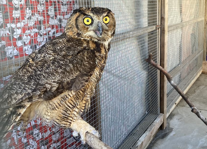
The great horned owl, Bubo virginianus, is a common patient seen at the Wildlife Medical Clinic. This species is found throughout most of North America and is easily identified by its “horns” which are actually tufts of feathers. Great horned owls are permanent residents of Illinois and can be found in a variety of habitats. This species is also known for its stereotypical “hoo-h’HOO hoo hoo” call, which is commonly heard in movies and on television shows.
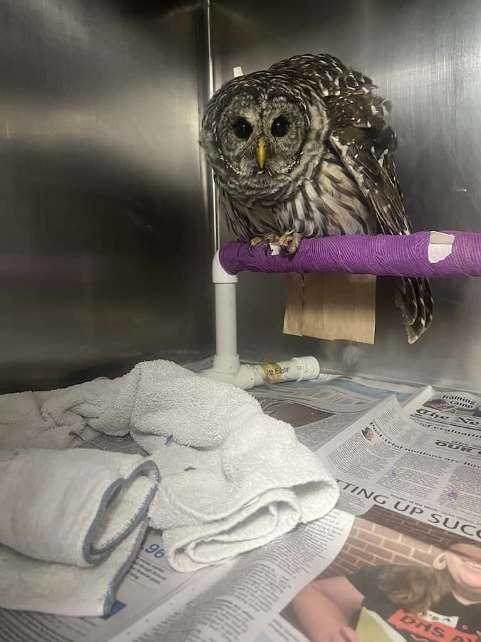
In the middle of August, a juvenile great horned owl was brought to the Wildlife Medical Clinic by a good Samaritan. The owl had been in the finders yard for a few days and was seen taking short flights but would fall out of the trees upon landing. The finder also noticed that the young bird was not using its right leg at all, and it seemed to be dangling from the joint. During the physical exam, the patient was found to have a proximal fracture of the right femur. The patient was also found to have external parasites, blood parasites, anemia, and was extremely underweight. Shortly after the initial exam, the patient had radiographs performed to confirm the fracture location and to evaluate whether surgery would be an option to repair the fracture. Upon review of the radiographs, it was determined that fracture repair was possible and had great potential to be successful, so the patient was taken straight to surgery.
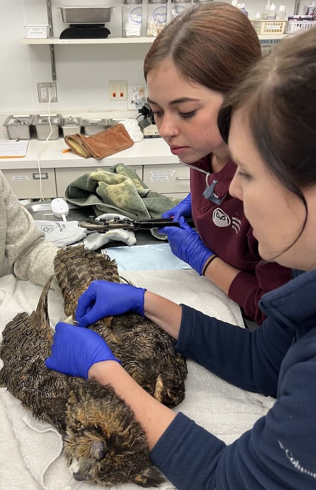
During the surgery pins were placed in the bone to stabilize the fracture and allow for the fractured pieces to be joined back together. Over the next six and a half weeks the patient was placed on cage rest while the bone healed. During this time, the patient was placed on antibiotics to help prevent infection, as well as medications to control pain and decrease inflammation or swelling. The patient was also treated for its parasites, which in turn treated the anemia the patient presented with. This period of rest also allowed the patient to gain weight and return to a healthy body condition. The patient’s surgical site was monitored daily for signs of healing and was kept clean by the volunteers. During the last week of cage rest, radiographs were repeated and confirmed that the patient’s fracture was healing properly and the surgical implant holding the bones in the correct position was ready to be destabilized. Destabilization essentially allows for the bone to slowly get used to supporting the weight of the patient.
When the pins were removed there was slightly more exudate than what would normally be expected. The material was cultured and sent to the Veterinary Diagnostic Laboratory at the University of Illinois to be examined by pathologists. The sample came back as an antibiotic resistant bacterium, staphylococcus epidermis. The bacteria was tested against different antibiotics until one was found to be effective against it. This type of infection is known as osteomyelitis, or a bone infection. Osteomyelitis happens infrequently but is associated with surgical repairs using pins in raptors. The patient was put on a course of twice daily antibiotics, and pain medications.
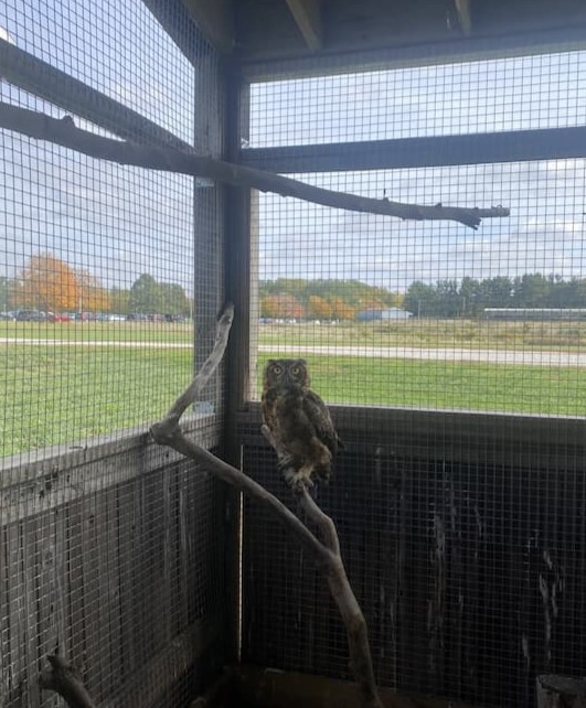
During this time, the owl was also moved to a larger enclosure in our flight cage, for further monitoring. This allows the doctors to confirm that the bone is continuing to heal properly and that the patient can use the limb correctly. This also allows for the patient to get used to using its leg and rebuild any muscle that may have deteriorated during the healing process. This is especially important because owls are birds of prey, which use their legs and talons to capture the food they eat. Owls also nest and perch in trees, which allow them to stalk their prey and stay safe from predators. Proper limb function is needed to keep the bird steady in the tree, and to catch their prey silently.
After three weeks of antibiotics and pain management the medications were discontinued, and the patient was monitored for any signs of pain, discomfort, or improper limb use. During this observational period the patient showed no clinical signs of infection or pain. Radiographs were also repeated and showed no evidence of damage to the bone from the infection. Upon further examination the patient was deemed ready for transfer to a rehabber to build up strength and sharpen its skills before being released to the wild. This is an important success story as Osteomyelitis is a rare surgical complication that can lead to the downfall of a patient. Luckily, the infection was caught early, and the patient responded well to the course of antibiotics giving him a chance to return to the wild, where he belongs!
Written by Maya, Class of 2026