Should You Feed Wildlife?

At the Wildlife Medical Clinic, we receive many phone calls from caring members of the community asking if they should be feeding healthy and/or injured wildlife. We truly appreciate when citizens care enough about our native wildlife to reach out and ask these commonly asked questions. The short answer is no, you should not feed wild animals. Although we understand that this gesture comes from a place of love for our wildlife species, it can cause harm to them and provide hazards to humans, as well.

That being said, let’s get into some specific examples of commonly fed wildlife.

Squirrels
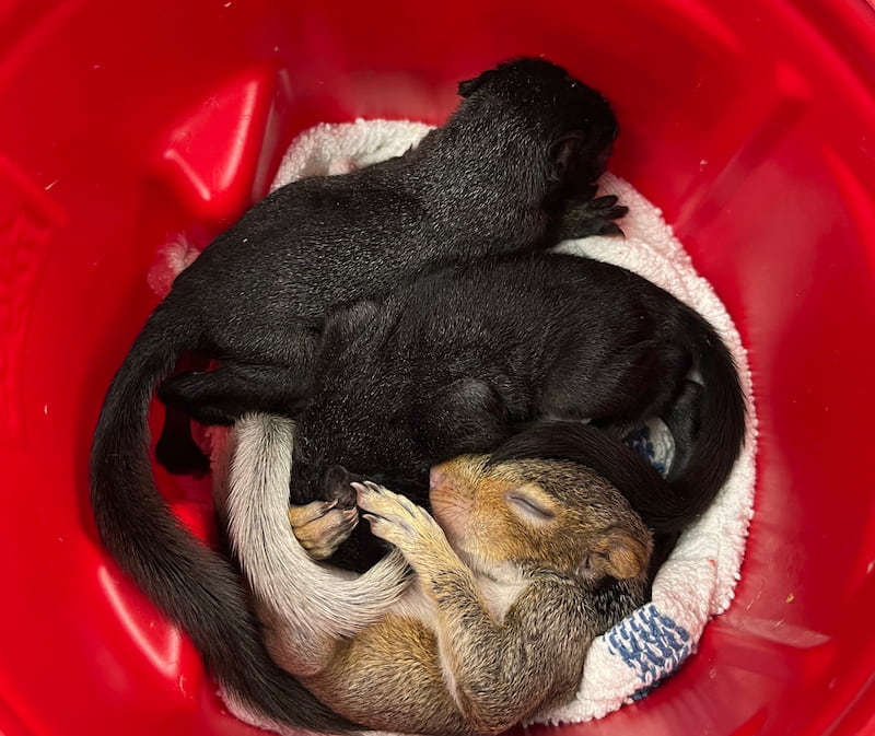
Small plastic container with three baby squirrels curled up asleep.

Squirrels are a common species of wildlife that we see frequently, even in urban areas! It is common for people to leave food out for the squirrels that live nearby, but let’s rethink that. Human food is often not nutritionally appropriate for their needs and can even cause illness when consumed. Not only can it be bad for their overall health, but it trains the squirrel to stop hunting and foraging for food. Then, if you forget to feed them once you’ve already started, or if you are away from your home for a period of time, these squirrels are unable to find food for themselves, which can lead to starvation. Squirrels that are being fed by humans often lose their fear of us and are therefore more likely to show aggression, which could result in biting a person. There are a multitude of human medical concerns that arise when bitten by a wild animal. To ensure your safety, your fellow neighbor’s safety, and the squirrel’s safety, please refrain from feeding them.

Raccoons
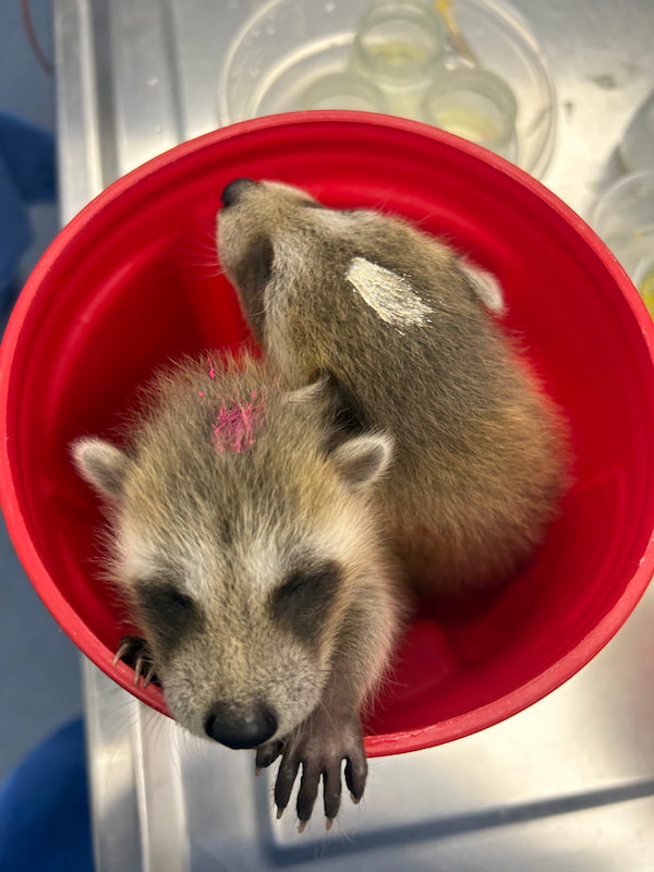
Two baby raccoon patients in a bucket.

For similar reasons to that of the squirrel, we also do not recommend feeding raccoons. When feeding raccoons, it can cause them to congregate in higher-than-normal numbers at your home. This upsets the balance of the local ecosystem and can increase competition for food. Thus, inducing more aggressive behaviors between raccoons, which can lead to conflict with humans. Raccoons are a common rabies vector species, and if bitten it would be considered a medical emergency. You should seek medical attention immediately or even go to the emergency room.

Birds

Bird feeders are a common route for feeding wildlife. One of the problems with using bird feeders is that it can aid in the spread of disease between birds. It can also lead to a higher rate of predation because there is now an influx of birds going to the same spot, on a predictable schedule. As bird feeders are usually next to or near windows, it can also increase the likelihood for window strikes, which can cause injury or even death to birds. Feeding birds has also been shown to change the behavior and health status of the bird.

What about Hummingbird Feeders?

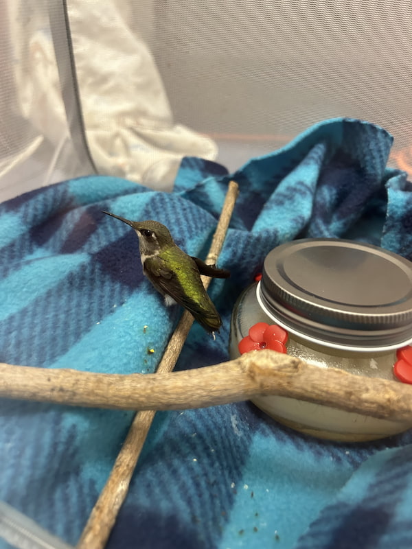
Hummingbirds are another species where there is a commercially available diet that people purchase to feed them. However, if regular cleaning is not completed, the nectar that is used to fill hummingbird feeders can easily become contaminated with various molds, bacteria, bugs, and fungus. This can cause illness and even death to the hummingbirds that visit those feeders.

Healthy Wildlife Should Not Need Help Finding Food

To ensure the health, safety, and protection of our native wildlife, please refrain from interfering in their natural behaviors by feeding them. We cannot match the same diet that these animals would be foraging for themselves in the wild and most commercially available diets marketed for feeding wildlife are not nutritionally balanced. As a licensed rehabilitator, we do have access to more nutritionally sound diets backed by research to help support these animals while they are in our care. We are also trained to take very strict precautions when overseeing these patients to ensure their safety and the safety of our staff. But the goal of our clinic is to safely rehabilitate wildlife and release them to their natural habitats as soon as possible.

How to Safely Interact with our Native Wildlife

As adorable as wildlife is, they are wild for a reason and do not need our intervention with food. However, there are some ways that you can still enjoy the beauty of our wildlife! For example, bird walks are a wonderful way to view different native avian species from a far. Also, cameras outside your home are a cool and interactive way to view your wildlife visitors!

Thank you for caring and protecting our native wildlife!

Written by Maddie, Class of 2027
HPAI – Best Practices

HPAI and Public Health

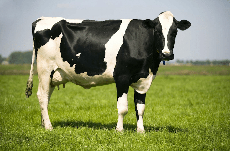
Highly pathogenic avian influenza, commonly referred to as HPAI, has become a common topic in conversation today. This may be attributed to the virus being detected in animals other than poultry or wild birds, including various mammalian predators and dairy cows, as well as several large avian mortality events attributable to the virus.

Avian influenza had previously been undetected in dairy cattle, but a discovery in 2024 identified the virus on certain farms. While the risk to public health is low at this point, preventing exposure is imperative in reducing opportunities for the virus to mutate. 

Preventing Infection

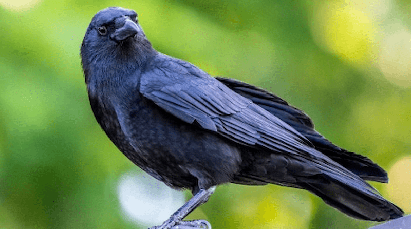
Picture of a crow for HawkTalk

The IDNR and IDPH urge the public to avoid physical contact with any waterfowl or any wild birds showing signs of illness. Pets can be infected by contact with carcasses, respiratory secretions, or droppings from infected animals, so it is important to keep your pets indoors or leashed and ensure they do not approach any sick or deceased animals.

Pets and humans can also be infected if consuming underdone infected poultry or raw milk from an infected cow. Being diligent to properly cook your food and avoid feeding your pet raw food or milk can prevent infection. If you have backyard poultry, keep them in a covered enclosure to prevent wild bird feces from dropping into the enclosure. Additionally, change your clothes and shoes before interacting with your poultry, and keep rodents and wildlife away from their food and water.

Deceased Wildlife

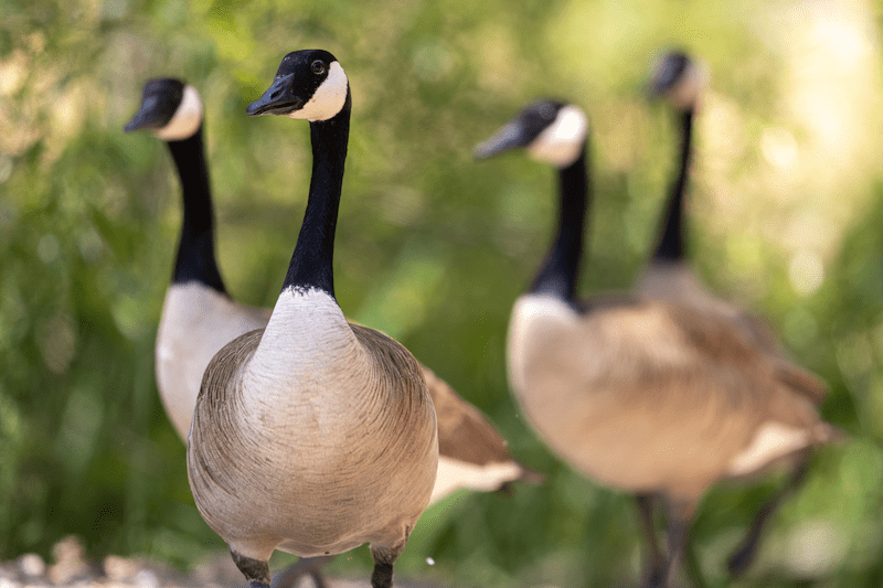
Canada Geese for HPAI HawkTalk

It is encouraged to avoid handling bird carcasses, but in the event carcasses must be removed from your property, there are steps you can take to protect yourself. If you come across a die-off of five or more birds you should contact your IDNR district wildlife biologist. When handling a deceased bird, you need to protect yourself by wearing the correct equipment. This includes waterproof gloves, safety goggles, an N95 mask, a hair covering, shoe covers, and a disposable gown or coveralls to protect your clothes.

Once you have your protective equipment on, you can dispose of the carcasses in a secure plastic bag and double bag the carcasses. Disinfect the outside of the bag with a disinfectant labelled for the flu virus and place it in a secure garbage can where animals will not get to the carcasses. When you are finished, carefully remove the protective equipment without contaminating yourself or clothes, double bag the equipment and again disinfect the outside of the bag and dispose of it in a waste container.

If you are unsure of how to remove your protective equipment, the CDC has information on how to safely do so. Make sure to wash your hands thoroughly afterward and avoid touching your face during the process. 

Monitor for Exposure 

If you have come into contact with a sick or deceased bird, even if disposing of a carcass with the correct equipment, you should contact your local health department, who will assess your exposure and help with symptom monitoring. Monitor yourself for flu-like symptoms after exposure and contact your health department if you develop symptoms up to 14 days after exposure. If you are experiencing symptoms and suspect you were exposed, do not show up to your healthcare provider without contacting them and the health department. 

References:

Centers for Disease Control and Prevention. (n.d.). Interim guidance for employers to reduce exposure to avian influenza A viruses for people working with animals. Centers for Disease Control and Prevention. https://www.cdc.gov/bird-flu/prevention/worker-protection-ppe.html

Press release. Illinois Department of Natural Resources. (n.d.). https://dnr.illinois.gov/press-release.30768.html 

How do I dispose of Dead Birds on my property (5 or more)?. Safe Dead Bird Disposal (5 or more Birds). (n.d.). https://dph.illinois.gov/topics-services/diseases-and-conditions/respiratory-disease/diseases/influenza/h5n1/dead-bird-disposal-5-or-more.html

Is the highly pathogenic avian influenza a threat to pets?. Veterinary Medicine at Illinois. https://vetmed.illinois.edu/wildlife/2025/01/09/is-the-highly-pathogenic-avian-influenza-a-threat-to-pets/

Written By: Alyssa, Class of 2028

That Bites!

Bite Wounds

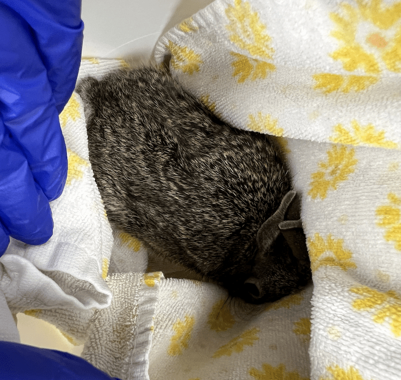
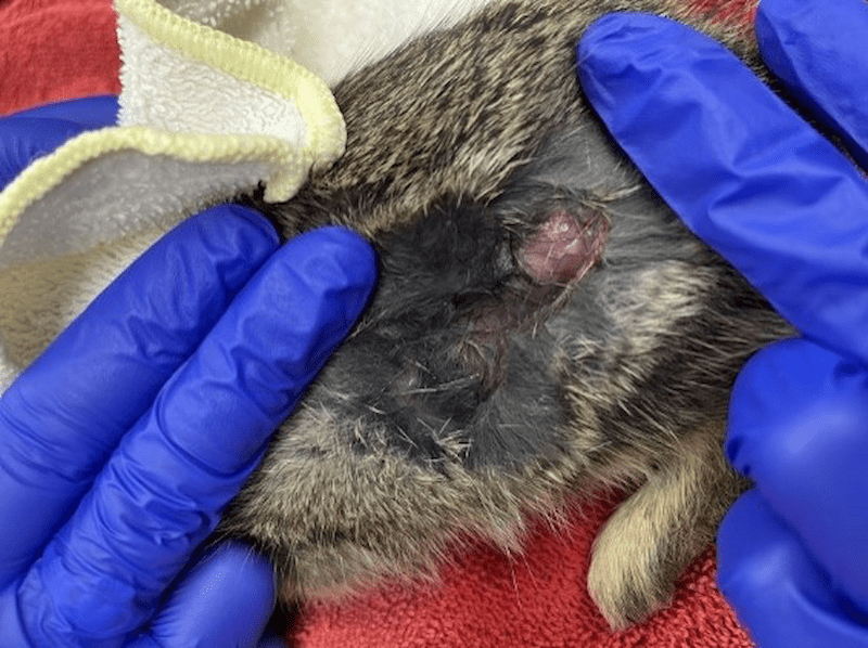
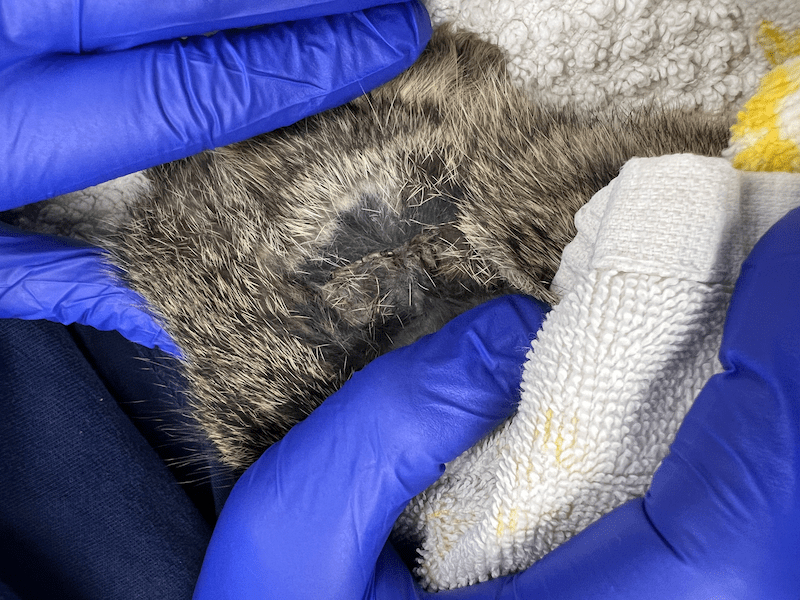
Initial presentation of an Eastern Cottontail with a bite wound from a cat

The Wildlife Medical Clinic has indeed seen it all in terms of injuries from human, domestic animal, and wildlife sources. Whether it be an accident or by instinct, all injuries, big and small, can become lethal to an animal if not adequately treated. One source in particular can be silent but deadly, wounds from a cat’s mouth.

A wound from a cat bite can harbor up to thirteen bacterial species from feline saliva. Most bites can host a mixture of multiple aerobic and anaerobic bacteria that can worsen puncture wounds and prevent healing or lead to sepsis. Several studies discovered that Pasteurella multocida is the most common pathogen within cat bites and is known to accelerate infections. At the Wildlife Medical Clinic, bite wounds are treated immediately with antibiotics and monitored very closely for any sign of infection that can indicate a difference between a healthy release and sepsis.

Case Correlation

The Wildlife Medical Clinic recently treated a juvenile eastern cottontail after it was bitten by a cat. Upon examination, a large 1.5 inch by .5-inch de-gloving, which is a wound where skin is pulled off the underlying muscle and tissue. Also, a wound was found over the left stifle (knee), but it did not appear to enter the abdominal cavity.

Because of the very thin and friable nature of wild cottontail skin (imagine wet tissue paper), it was decided that any attempt to clip, clean, or suture the wound could cause further damage. Additionally, bite wounds should not be sutured closed, as that can trap dangerous bacteria under the skin. A simple flush of the wound was performed to remove excess debris.

A few other small abrasions were noticed but ultimately were of no concern. The eastern cottontail was ambulating (moving around) appropriately, was of a healthy weight, and had only mild dehydration. We decided that the best course of treatment was to provide fluids, pain medication, and antibiotics. To minimize stress and worsening of the wound, we were hands off as much as we could be with this patient. 

The Outcome

Young rabbits can heal their wounds with impressive speed, and several days later, the wound began to show signs of healing. With no signs of infection or pain, antibiotics and pain medications were discontinued from daily treatment with the plan to continue to monitor. Within eight days, the wound was fully scabbed and posed no threat to normal movement. The eastern cottontail was of healthy weight and was cleared for release! Another successful and rewarding case at the Wildlife Medical Clinic at the University of Illinois!

References:

Talan, D. A., Citron, D. M., Abrahamian, F. M., Moran, G. J., & Goldstein, E. J. C. (1999). Bacteriologic analysis of Infected Dog and Cat bites. New England Journal of Medicine, 340(2), 85–92. https://doi.org/10.1056/nejm199901143400202 

Dire, D. J. (1991). Cat bite wounds: Risk factors for infection. Annals of Emergency Medicine, 20(9), 973–979. https://doi.org/10.1016/s0196-0644(05)82975-0

Written by: Madison, Class of 2027

Nutritional Secondary Hyperparathyroidism

What is Nutritional Secondary Hyperparathyroidism (NHSP)?

Nutritional secondary hyperparathyroidism (NSHP) is unfortunately a common type of metabolic bone disease that can occur in any animal but is most often seen in reptiles and amphibians. This condition most often occurs in captivity when an animal is not receiving the proper husbandry. NSHP can occur when the animal’s diet does not have enough calcium, or vitamin D3, or in the case of many reptiles when they do not receive enough UVB (ultraviolet B) light. Vitamin D3 is required by the body for calcium absorption, so as a result the parathyroid gland, which is found in the neck with the thyroid, begins to produce more parathyroid hormone (PTH). The primary function of the parathyroid gland is to tightly maintain calcium levels and keep them balanced. As a result of the decreased calcium and lack of vitamin D3 the secreted PTH stimulates osteoclasts in the bones, where most calcium is found, to begin resorbing the bones breaking them down causing the calcium to be released back into the bloodstream. Calcium is extremely important throughout the body where it is required for nerve transmission, muscle function, blood clotting and a multitude of other vital bodily functions. While the calcium needs to be in the blood it does so at the expense of the bones.

Why do we see NSHP in Wildlife?

When the bones are broken down to provide calcium it causes them to lose density making them more susceptible to injury. Additionally, there are developmental deficiencies associated with growth that can occur too. All of these make the lives of animals very difficult. NSHP is not as prevalent in the wild, because the animals tend to get enough sun and eat a proper diet. There are two main reasons we see this, first is when people have a pet and they don’t do proper research on housing and diet requirements and therefore don’t meet the needs of their pet. The second reason we see this is due to well-meaning individuals finding wildlife and then proceeding to care for them or keep them in captivity instead of bringing them to a licensed rehabilitator. Wildlife requires specialized diets and housing that are hard to meet in captivity and while people try their best, they often fail to meet the needs of these animals and don’t realize that for most wildlife, permits are needed to care for them, so keeping them in captivity is actually illegal if you don’t possess the proper permits. To further illustrate the effects of NSHP, we will review two cases we have recently seen in the Wildlife Medical Clinic.

Virginia Opossum Case

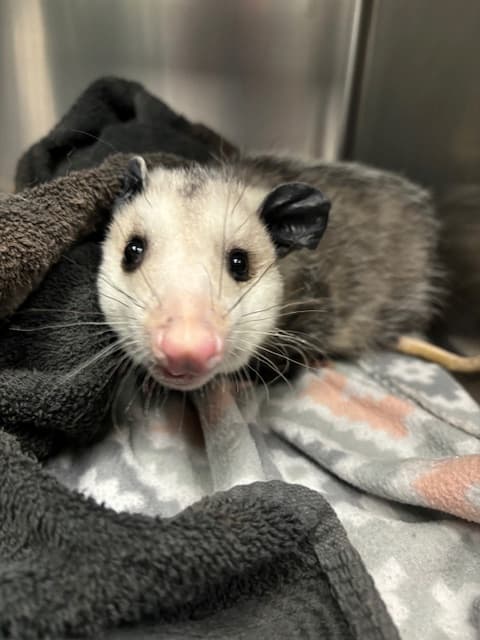
Virginia Opossum with non-symmetrical face due to metabolic bone disease.

On September 9, 2024, we received a juvenile male Virginia opossum that a finder found in their yard struggling to walk. Upon exam he struggled to walk and lacked coordination in his back legs. Additionally, his face was swollen and asymmetrical. Due to the possible orthopedic issue and significance of ataxia we opted to take radiographs.

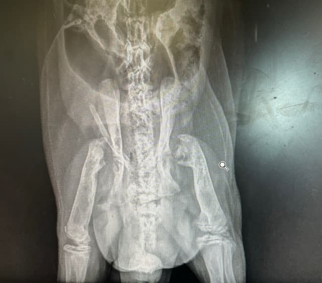
Virginia opossum x-ray.

The radiographs showed that the opossum had a decrease in bone density, and severe malformations of the long bones primarily at the femoral head which is the part of the femur that connects to the hip. They were so degenerated that they appeared to be absent. All the joints appeared to be affected in some way, but that was the most notable location. These changes to the bones and the facial asymmetry led to our diagnosis of nutritional secondary hyperparathyroidism. Our belief is that he was kept as a pet for a significant amount of time when he was younger by someone who did not have the proper knowledge or credentials and then he was released. He also had a very high body condition score indicating he ate well, which would not have been possible on his own, in the wild, due to his musculoskeletal abnormalities. Even though he ate well, he did not receive the proper nutrients needed for bone formation and maintenance. Unfortunately, due to the severity of the malformations and the fact that multiple joints were affected the most compassionate care we could provide was humane euthanasia. He likely had arthritis, which is painful and overall had a poor prognosis for surviving in the wild as the damage done was irreversible.

Common Snapping Turtle Case

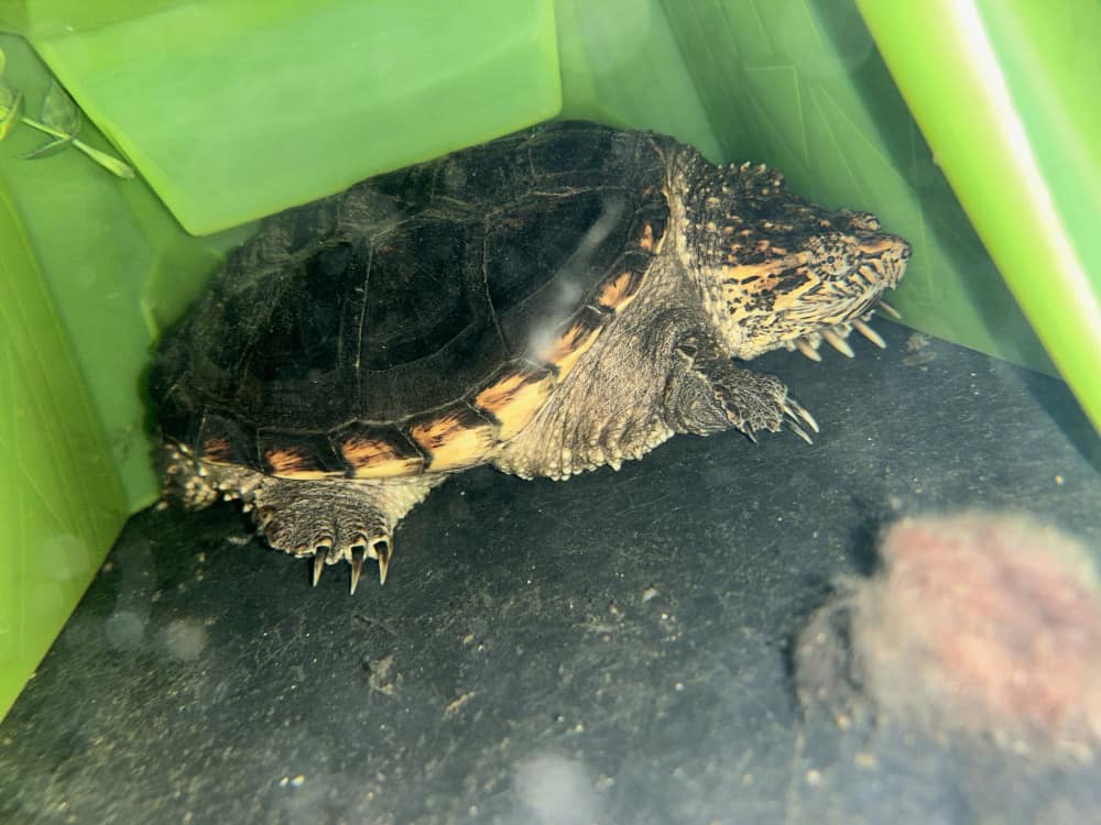
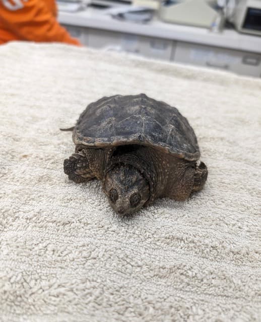
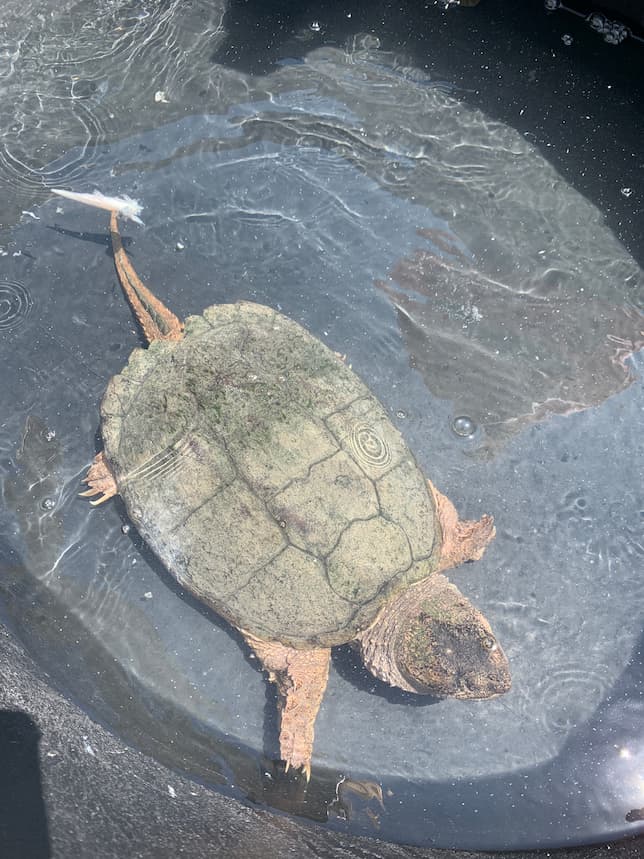
Common snapping turtle on exam table for initial exam after arriving to the Wildlife Medical Clinic.

While the Virginia opossum did not have the outcome we hoped for, our next patient, a common snapping turtle, has a more positive outcome. On November 11, 2023, we received a juvenile male common snapping turtle after it was found abandoned in a vacated apartment where he was owned for around two years. He was found dry docked and severely dehydrated. Upon physical exam he was severely dehydrated, his shell appeared too small for his body and was soft and flat. Additionally, he had signs of stunted growth. All of this points to NSHP, primarily the softening of the shell and stunted growth.

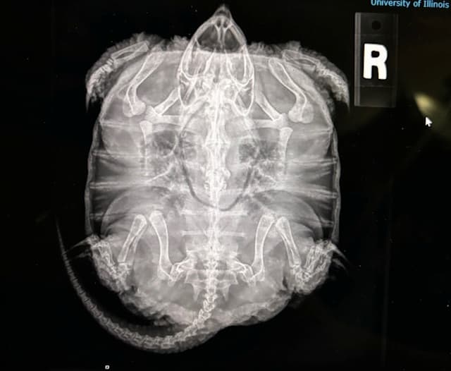
Common Snapping turtle x-ray

This was confirmed through radiographs, which showed decreased bone density and stunted growth Fortunately, unlike the opossum the damage was reversible. He was put on a proper diet along with consistent UVB and heat and now just over a year later he has grown a considerable amount and his body condition has greatly improved. While this is great news, he still is not releasable due to his friendly nature. Because of the time he spent in captivity, he is habituated to people. We have been working on finding him a permanent home and have received interest from an out of state nature center. However, if for some reason this home doesn’t work out, we will keep looking.

Lessons Learned from these Cases

These two cases illustrate the importance of nutrition and proper care for animals. If you want to own a pet, make sure you do the proper research and have everything set up and ready for them before you bring them home. Picking out a pet should not just be done on a whim. They are living creatures that require care and work. The snapping turtle shows the importance of doing research and being committed to owning a pet. Obviously, we don’t know the circumstances of his abandonment, but this is unfortunately an all too common occurrence when it comes to reptiles. Reptiles are unique in their dietary and husbandry needs, which need to be known by the owner, or else serious health issues like metabolic diseases can arise. The case of the Virginia opossum illustrates the importance of knowing who to contact, and only taking care of wildlife if you have the proper knowledge, credentials, and permits. The WMC is built on the idea of us working with the public to better the quality of life for wildlife. If you see an animal that you think needs help, don’t try to keep it and care for it on your own, because it can put you and the animal in danger. Contact a licensed rehabilitator or reach out to the Wildlife Medical Clinic and we can help provide guidance on what to do and who to contact. We hope these cases illustrated how important animal nutrition and husbandry is, why it is best to leave wildlife in the wild, and if help is needed to please contact a licensed rehabilitator.

Written By: Dylan, Class of 2026

Preparing for Spring Migration

Spring migration is a magical time when birds return to their breeding grounds after a long winter. However, this journey can be perilous, with various challenges along the way. Dr. Stephany Lewis DVM CWR DABVP (Avian Practice), and Sarah Hollander, Wildlife Ambassador Coordinator will share insights on how to support migrating birds and other wildlife during their stopovers in your yard. 

https://www.wcia.com/ciliving-tv/wildlife-medical-center-is-preparing-for-spring-migration/

American Crow and The West Nile Virus 

American crow (Corvus brachyrhynchos), one of the most intelligent bird species in North America. They are very social birds and sleep in a communal roost together in the winter! They thrive by working collectively and using their intelligence to catch or steal prey and other food items. Yet, this unique species has faced the threat of West Nile Virus since 1999 when the outbreak began in New York City. According to the data, this disease has caused a 45% regional decline in crow populations.  

West Nile Virus (WNV) is a vector-borne disease that is spread by mosquitoes. In a temperate climate, WNV is prevalent in late summer and early fall. It can infect a wide range of species, from birds to humans. Mosquitoes play a critical role in the transmission, as they are the vector. Meaning they are the intermediary. They get the virus from the blood of one host, for example an infected bird, and then when they bite another species like a human, they pass the disease through the bite. Transmission has also been documented from bird-to-bird. This usually occurs when a bird consumes another bird that is infected with WNV or when water tainted with feces and saliva from an infected bird is drank.    

Over 250 bird species have been documented as WNV-positive cases. Within these species, the corvids (crow, raven, and jay) have the highest susceptibility to WNV in North America. Many studies have investigated this phenomenon and found correlations between human WNV cases, mosquito transmission pathways, and crow death from WNV. Rapid urbanization significantly impacted the speed of virus transmission in wildlife.  

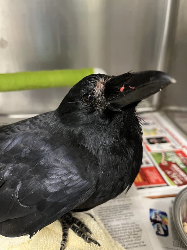
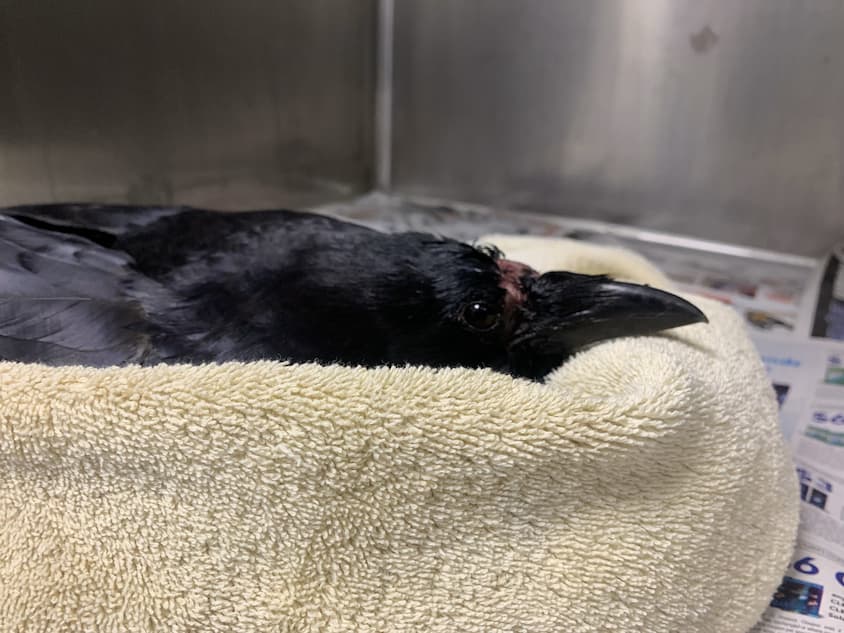
A crow laying on a blanket

West Nile Virus in birds primarily attacks the central nervous system. Therefore, some of the symptoms an infected bird displays include a wide range of neurologic signs, such as a head tilt, lethargy, weakness, and loss of coordination. Many cases that come across the Wildlife Medical Clinic also show a great degree of emaciation, where patients are significantly underweight and are unable to fly. Once the crow is infected with the virus, it almost always indicates a grave prognosis. Studies have shown that most infected crows die within three weeks of contracting the virus. And testing for the disease will likely occur post-mortem through tissue samples from the heart, brain, and kidneys or via oral swabs.  

The success rate of recovery is incredibly low in crows, since no known established treatments are available to clear the viral infection. Most cases rely on the animal to clear the virus and recover with supportive care. And the mortality rate can be as high as 100%. However, some breakthrough discoveries were made in 2022 by the Cornell College of Veterinary Medicine – where their wildlife experts successfully treated five infected crows with fluids, anti-inflammatory drugs, anti-fungal drugs, and anti-parasitic drugs. This treatment successfully allowed these wildlife experts to release the five American crows that were previously ill with WNV. The discovery was linked to potential immune system adaptation in the crow or the shifting of the virus’s behavior. While this treatment was successful, cases in general still pose a grave prognosis for crows and research continues in order to better understand this disease dynamic. We are hopeful that a treatment will become available in the future that will help our Crow friends fight off this deadly virus.

Written by YiYing, Class of 2026

References

Wheeler, S.S., Taff, C.C., Reisen, W.K. et al. Mosquito blood-feeding patterns and nesting behavior of American crows, an amplifying host of West Nile virus. Parasites Vectors 14, 331 (2021). https://doi.org/10.1186/s13071-021-04827-x 

Yaremych SA, Warner RE, Mankin PC, Brawn JD, Raim A, Novak R. West Nile virus and high death rate in American crows. Emerg Infect Dis. 2004 ;10(4):709-11. doi: 10.3201/eid1004.030499. PMID: 15200865; PMCID: PMC3323091. 

Johns Hopkins Medicine. West Nile virus (n.d.). https://www.hopkinsmedicine.org/health/conditions-and-diseases/west-nile-virus 

Wildlife Futures Team. West Nile Virus. PennVet – Wildlife Futures Program. (2021). https://www.vet.upenn.edu/research/centers-laboratories/research-initiatives/wildlife-futures-program/our-research/fact-sheet-detail/west-nile-virus#:~:text=Birds%20with%20West%20Nile%20virus,will%20die%20within%203%20weeks. 

Cordova, M.G., Cornell documents first crows to survive deadly West Nile virus. Cornell Chronicle. (2022). https://news.cornell.edu/stories/2022/06/cornell-documents-first-crows-survive-deadly-west-nile-virus 

Emerging Turtle Disease, Emydomyces Testavorans 

Emydomyces testavorans is a recently discovered fungus associated with freshwater habitats that causes shell disease in a wide variety of turtles and terrapins (Woodburn et al 2019; Woodburn et al 2021). The fungus has been found in both wild and human cared for turtles and has been identified in individuals with active lesions and those with no evidence of disease.

E. testavorans was originally discovered in animals housed in captive care but recent studies found that it has the ability to infect and cause disease in wild populations as well (Lambert, MR, 2021).

Once established, lesions gradually eat away at the shell, progressing through the bone which also exposes turtles to infections. These lesions can have a wide variety of presentations from ulcerations of the shell, pitted depressions, discoloration and flaking of the shell, increased pliability of the shell, nodular masses within the shell or abdominal cavity, and cystic nodules. 

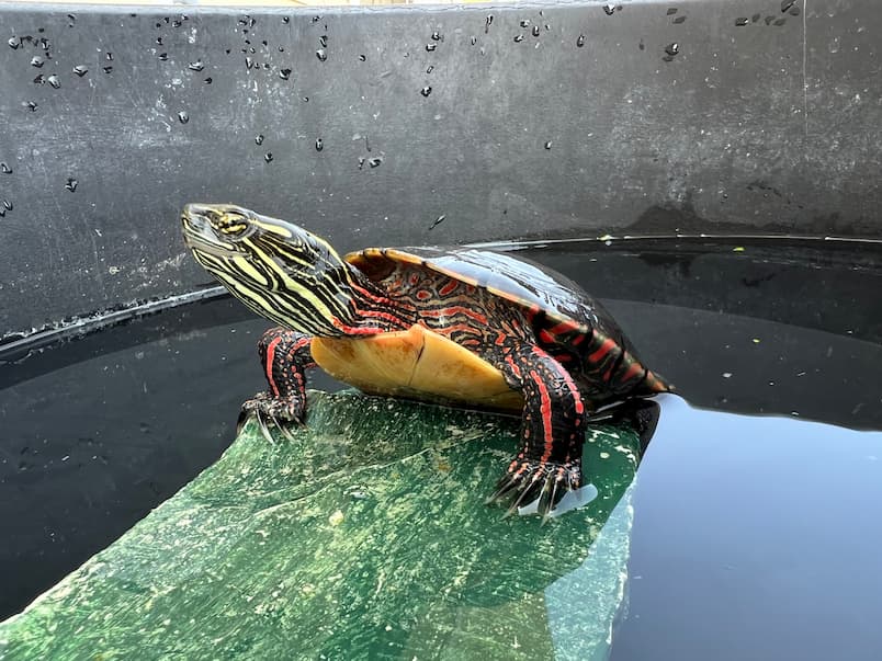
A turtle laying in water

The presence or absence of E. testavorans can be detected within shell tissues through polymerase chain reaction (PCR) run from swabs or scrapes of shell lesions and/or fresh or frozen tissue.  Histopathology and necropsy can also be helpful in identifying whether an infection is present through stained shell tissues in the lab. 

The recommended treatment for shell disease secondary to E. testavorans is still being investigated but nebulization of the anti-fungal terbinafine once daily for 28 days has been shown to be helpful, especially in addition to antifungal medication given over a set period of time and repeated in set intervals to help prevent reoccurrence. This is also known as pulse antifungal therapy. Other forms of treatment have included debridement of necrotic shell, packing lesions with terbinafine powder, and covering them with bone wax or cement. 

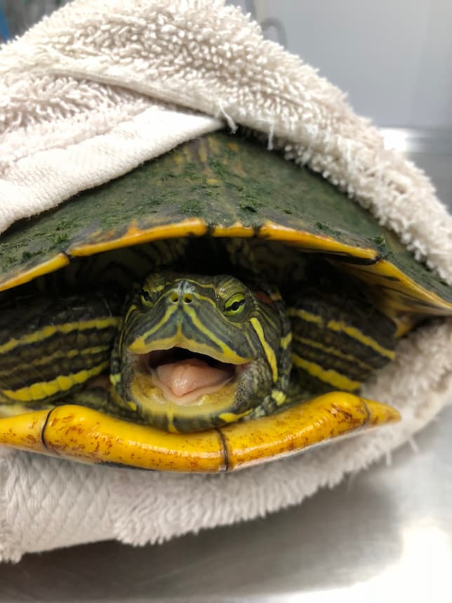
A turtle wrapped in a blanket

Unfortunately, shell disease is a common cause of illness and death in turtles and the underlying cause for why some turtles will develop more severe disease remains unknown. Turtles can either be asymptomatic carriers or show active evidence of infection; however, those that show evidence of infection are more likely to succumb to severe, chronic disease of the shell and a large number of those severely infected turtles will die. 

Identifying the fungus is an important first step which allows us to better understand how the fungus spreads which can help us to develop health management strategies for impacted turtles, and ways to combat potential emerging disease. Having this knowledge provides us with not only the means to quickly diagnose patients and implement possible treatment options, but is also essential in assisting to develop proper handling and biosecurity measures as well. 

*Turtle images featured in this article are for visual design. They do not have the disease mentioned.

Written by Tori McCarthy, Affiliate Clinical Year DVM Student   

Wildlife Medical Clinic
John A. Coyne South Clinic
2100 South Goodwin Avenue
Urbana, IL 61801
217-244-1195