
LAB ACHIEVEMENTS
Decreased lymph node estrogen levels cause nonremitting progressive experimental autoimmune encephalomyelitis disease
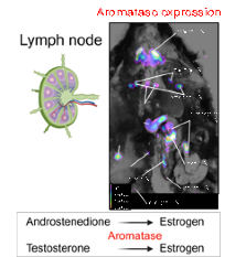
Estrogen plays a pivotal role in modulating immune responses. This study explored the expression of aromatase, the enzyme responsible for estrogen synthesis, in lymph nodes (LNs) and its potential role in the pathogenesis of MS using a mouse model. Targeted delivery of an aromatase inhibitor specifically to LNs induced an interferon-β-resistant experimental autoimmune encephalomyelitis (EAE) phenotype. Our findings underscore LNs as crucial sites of de novo 17β-estradiol production, which may contribute to non-remitting EAE phenotypes.
Read more at DOI: 10.1093/pnasnexus/pgaf010
This study was performed in collaboration with Dr. Jay Ko.
Star-Shaped Glatiramer Acetate Mitigates Pulmonary Dysfunction and Brain Neuroinflammation in a Murine Model of Cryptococcus-Associated IRIS.
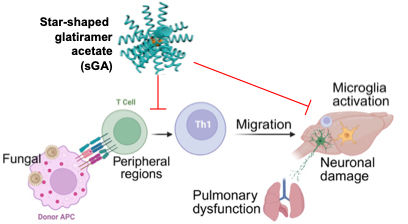
sGA exerts neuroprotection in Cryptococcus-associated immune reconstitution inflammatory syndrome (CIRIS) by limiting peripheral CD4+ T cell effector activity and suppressing CNS-resident immune activation. This study supports the use of sGA as a promising preclinical therapeutic candidate for C-IRIS and other Th1-mediated neuroinflammatory conditions.
Read more at DOI: 10.1101/2025.01.07.631707
This study was performed in collaboration with Drs. Song, Sharma, and Cheng.
T Cell Infiltration into the Brain Triggers Pulmonary Dysfunction in Murine Cryptococcus-associated IRIS.
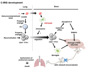 Cryptococcus-associated immune reconstitution inflammatory syndrome (C-IRIS) is a condition found in immunocompromised patients on antiretroviral therapy and characterized by numerous symptoms including pulmonary distress. Here, we use a mouse model to characterize the processes underlying this pulmonary dysfunction and find that infiltrated CD4+T cells into the brain trigger the injury of NTS neurons, which control pulmonary function. Thus, C-IRIS mice show severe pulmonary dysfunction and eventually mortality.
Read more at DOI: 10.1038/s41467-023-39518-x
This study was performed in collaboration with Drs. Takahashi and Sharma.
Estrogen receptor alpha signaling in dendritic cells modulates autoimmune disease phenotype in mice.
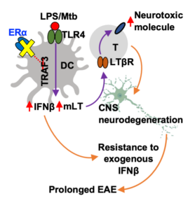
ERα signaling in dendritic cells suppresses the activation of neurotoxic CD4+ T cells and thereby modulates mouse autoimmune disease phenotypes. This effect is mediated by endogenous estrogen derived from extra-gonadal sources. Estrogen derived from extra-gonadal sites modulates phenotype severity in a mouse model of experimental autoimmune encephalomyelitis (EAE) dependent on ERα function in dendritic cells (DCs).
This study was performed in collaboration with Dr. Ko (UIUC).
Read More: https://doi.org/10.15252/embr.202154228
Astrocytes lure CXCR2-expressing CD4+ T cells to gray matter via TAK1-mediated chemokine production in a mouse model of multiple sclerosis
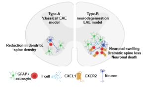 Multiple sclerosis (MS) is a chronic neurological disease characterized by demyelination and neuronal damage. While T cells are consistently detected in the gray matter of human MS samples, they are rarely seen in the gray matter of the most common mouse model of MS. Here, we show a modified mouse model that is characterized by a high degree of neuronal damage, T cell infiltration, and reactive gliosis in spinal cord gray matter. Using conditional knockout mice, we show that T cell migration to spinal cord gray matter depends on T cell expression of CXCR2 and astrocyte expression of TAK1-mediated chemokines such as CXCL1.
This study was performed in collaboration with Dr. Steelman (UIUC).
Read more: https://www.pnas.org/content/118/8/e2017213118.short
Early-life-trauma triggers interferon-β resistance and neurodegeneration in a multiple sclerosis model via downregulated β1-adrenergic signaling
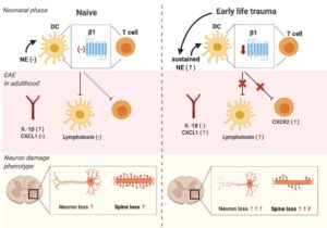
Environmental factors affect multiple sclerosis (MS) risk and disease development. Exposure to early life trauma (ELT) has been associated with higher relapse rates in MS patients; however, the underlying mechanisms are not well-defined. We show ELT induces mechanistic and phenotypical alterations during experimental autoimmune encephalitis (EAE). Altered EAE disease phenotype was attributed to ELT-induced chronic downregulation of β1 adrenergic receptors in dendritic cell and T lymphocyte. Our results indicate that ELT alters EAE phenotype via downregulation of β1 adrenergic signaling in immune cells.
Read more: https://www.nature.com/articles/s41467-020-20302-0
Immune regulation of CNS dysfunction in Cryptococcus-associated Immune Reconstitution Inflammatory Syndrome (C-IRIS)
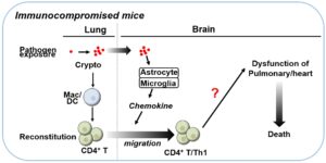
Immune reconstitution inflammatory syndrome (IRIS) is a pathological condition whereby a recovering immune system paradoxically worsens the patient’s condition by responding excessively to a previously acquired infection. IRIS has been reported in patients, who are recovering from an immunocompromised condition and pre-infected with fungi such as Cryptococcus (C-IRIS). We reported a mouse model that presents phenotypes similar to human C-IRIS symptoms, such as systemic upregulation of some inflammatory cytokines and brain edema. We are investigating how CNS-infiltrated CD4+T cells regulate neuronal function in the context of respiratory failure.
This study was performed in collaboration with Dr. Shinohara (Duke University).
Read more: https://www.frontiersin.org/articles/10.3389/fimmu.2020.529219/full
Development and improvement of treatment options for multiple sclerosis using EAE model
 Glatiramer acetate (GA) significantly reduces relapse rate and the appearance of lesions by magnetic resonance imaging in MS patients compared with placebos. Despite the broad use of GA in MS treatment, GA use is limited by tolerability issues due to the need for frequent injections. we reasoned that changing the structure of GA may alter its degradation and release profiles, thus achieving good efficacy while maintaining low-dose and less-frequent injections. We reported that star-shaped GA (sGA) exhibited much better performance than linear GA in treatment of experimental autoimmune encephalomyelitis (EAE). Even at a lower dosage with a single injection, sGA suppressed motor dysfunction and CNS demyelination in EAE mice.
This study was performed in collaboration with Dr. Cheng (UIUC).
Read more: https://pubs.rsc.org/en/content/articlehtml/2020/bm/d0bm00957a
Contribution of peripheral immune cells in CNS damage: Neutrophil CXCR2 activation by CXCL1 upregulates reactive oxygen species production but not CNS migration in EAE with severe neurodegeneration
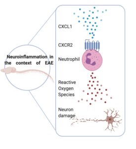 CXCR2 is thought to be the main receptor in regulating neutrophil chemotaxis and effector function during inflammation. CXCR2 signaling can be activated by receptor ligand CXCL1 which has been shown to be increased in MS patients. Our study provides evidence of neutrophil-driven neuronal swelling and synaptic loss that is dependent on CXCR2 signaling which is a key regulator in ROS production.
Read more: https://jneuroinflammation.biomedcentral.com/articles/10.1186/s12974-020-1730-y#Sec24
Development of super-resolution tool to visualize neuron morphology
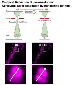
Metal‐based Golgi‐Cox (GC) staining is an established method used to visualize neurons with great morphological detail. GC stained samples are imaged routinely under transmitted light microscopy. This method is unable to yield information on the three‐dimensional structure of neurons. We show that the Confocal Reflection Super-Resolution (CRSR) technique can be used in conjunction to visualize both GC and immunofluorescence targets to create precise and improved three‐dimensional visualization and analysis.
This study was performed in collaboration with Dr. Sivaguru (UIUC).
Read more: https://onlinelibrary.wiley.com/doi/full/10.1111/jmi.12821