Case by Faith Ramsey, VM19
Triage and Presentation:
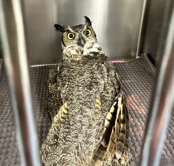
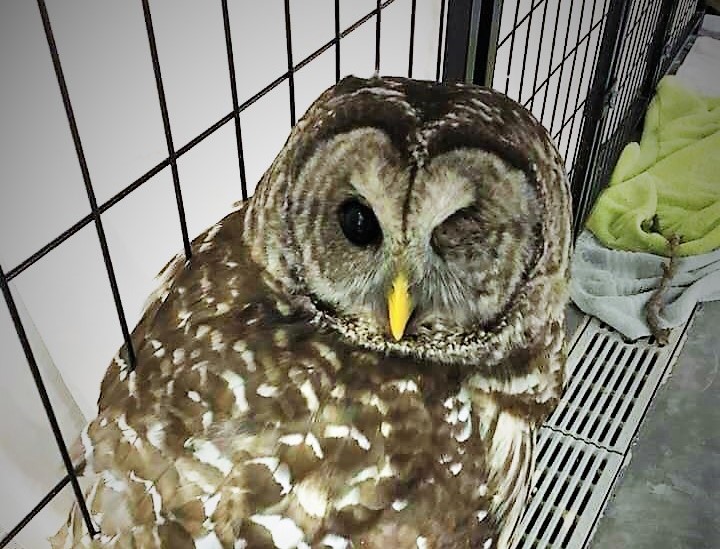
A Barred Owl was presented to the University of Illinois Veterinary Teaching Hospital Emergency Room in November 2017 for head trauma and hyphema (blood) in the left eye.
The following morning, a full physical exam was performed by a WMC. The patient was BAR and had a BCS of 2.5/5. The hyphema in the left eye covered the pupil and a Pupillary Light Reflex (PLR) could not be assessed in that eye. The owl’s sight was likely obscured in the left eye. PLR was noted to be normal in the right eye. Dried blood was noted on the medial aspect of the tarso-metatarsal (lower) region of the right leg. Ectoparasites (pigeon louse flies and smaller mites) were found to be crawling on the patient. All other systems were deemed normal.
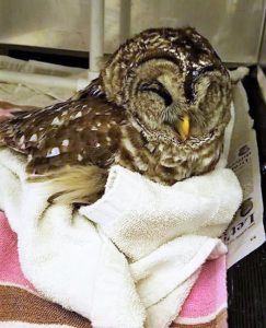
The team attempted to place an IV catheter unsuccessfully. Fluids with added B vitamins for energy were given subcutaneously, split between both inguinal regions. The patient was then wiped down with fly/mite spray to lessen the ectoparasite burden. The team calculated maintenance and replacement fluids. Medication calculations for pain, inflammation, and infection were placed on the treatment plan.
Treatments:
The following day, the Veterinary Teaching Hospital’s Ophthalmologists performed an ophthalmology examination. They determined the right eye did not have any changes that were significant enough to affect vision. The ophthalmologists confirmed hyphema of the left eye, and also noted fibrin formation in the anterior chamber. The fundus was obscured, and the globe was abnormally soft. An evisceration was quickly planned – this procedure removes the eye entirely (this is a valid option for owls, as they primarily use their incredible sense of hearing for hunting).
This patient decided it did not want to eat, so the team elected to forceps-feed twice a day. The fluids regimen was continued for three days. The bird’s left eye was assessed daily to make sure it would continue to be a good candidate for surgery.
Surgery:
The day of surgery arrived, and the patient looked ready! An IV catheter was placed for quick venous access during surgery. Feathers of the left periocular region were minimally plucked, and the eye was prepared aseptically. The surgery proceeded without incident. Because this was an evisceration, the lens and intraocular tissues were gently removed instead of a complete enucleation. This is due to the scleral bone being needed for proper hearing, so the animal can hunt once released. Recovery was uneventful.
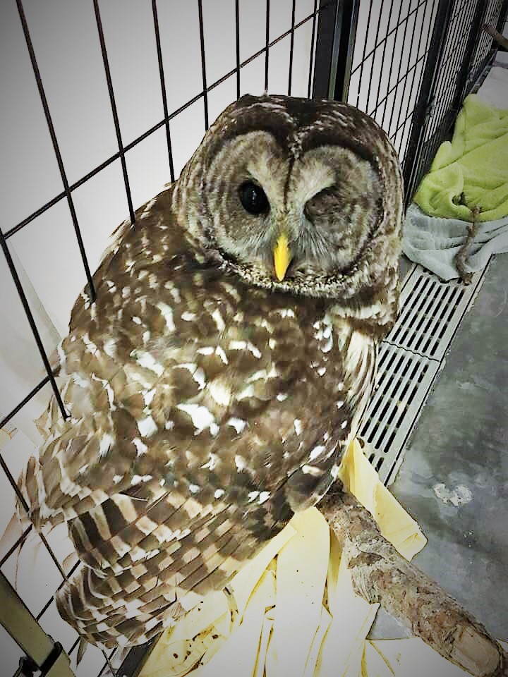
Post-Op Recovery:
Over the next few days, the patient was treated for pain and continued to be forceps-fed mice. Once healing was deemed to be going well, the patient was moved to a larger cage. After another few days, the patient began to eat thawed whole chicks on its own (volunteers learned it preferred chicks this over mice)! The patient was doing well enough that it was deemed releasable! On December 1st the patient was taken out to Douglas-Hart Nature Center in Matoon, IL, close to where it was originally found.