When a patient is brought to the Wildlife Medical Clinic, we have a multitude of options for diagnostic testing. Many of our patients undergo blood tests and radiographs (x-rays), but our options don’t stop there! Our Veterinary Diagnostic Laboratory is equipped to process any number of samples, including swabs, blood, and biopsied tissue. Having the expertise of laboratory technicians and pathologists means we can optimize the value of every sample we collect. Because we are in the Veterinary Teaching Hospital, we can also utilize equipment like ultrasound, CT (computed tomography), MRI (magnetic resonance imaging), and endoscopy. In addition to these modalities, we have the invaluable expertise of veterinarians trained to utilize them and eager to teach all of us students how to do the same. Here’s a little bit of information that helps us decide when to utilize different diagnostic tools and tests.
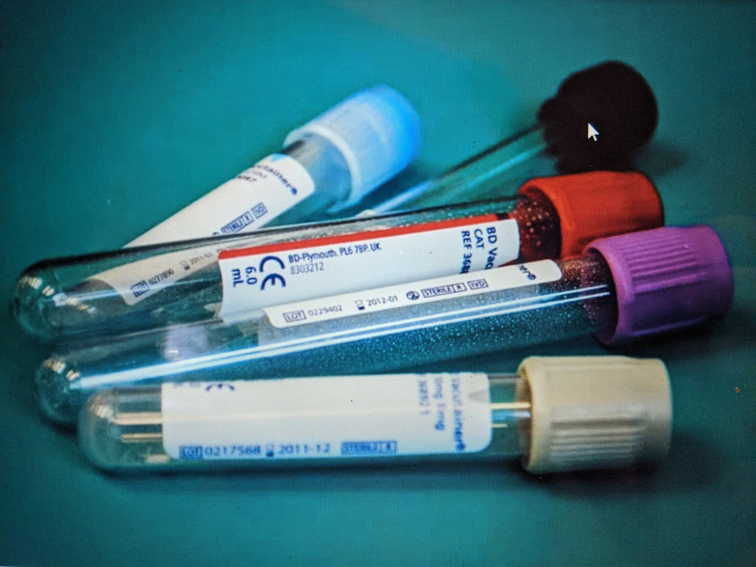
Blood tests can give us clues into the overall health of our patient. These samples can offer a general indicator of health or illness, like showing evidence of blood loss or dehydration. Each of the clues offered from these initial tests can lead us to a treatment or help guide our next diagnostic steps. We can also look at indicators for damage of specific organs, such as the liver or kidney. Blood samples can further be utilized to identify specific causes of disease, such as identifying blood parasites on a microscope slide or the presence of antibodies to a specific disease in the serum.
When it comes to orthopedic (bone) injuries, radiographs (x-rays) are often our go-to diagnostic test. Taking radiographs is efficient and gives us detailed information about what is happening inside our patient. The speed of the procedure can be important, as our patients must be sedated or anesthetized to get appropriate images. If a patient is very debilitated, prolonged anesthesia can be dangerous. Radiographs are a quick and appropriate way for us to learn the extent of a fracture and to help us determine if and how we can repair it.
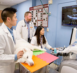
Ultrasound is useful in offering real-time image of specific organs. Patients don’t always need to be sedated or anesthetized to utilize an ultrasound. We can get an idea if there is fluid accumulating somewhere it shouldn’t (like the chest) and identify lesions in an organ. Because it is so fast, we can quickly identify signs of internal trauma and make informed decisions based on the prognosis for the type of injury. Unlike radiographs, ultrasound cannot “see through” bone, so it’s not as helpful if you’re trying to evaluate an animal’s bones.
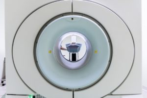
CT and MRI are similar in that they give us detailed images of the patient and are not used in the WMC as routinely as radiographs. In the case of both MRI and CT, the patient must stay completely still and is anesthetized for the procedure. These tests are performed by laying the patient on a table and rolling them into the machine. A CT takes radiographs at several different angles to create a 3-D model on a computer program. The model can then be rotated, looked at in sections, or even printed into a 3-D version. This can be useful if there is a difficult to repair fracture – surgeons can complete the repair utilizing a model before doing it on the actual patient! MRI uses magnets instead of radiation and can also yield detailed images of what is happening in the patient. An MRI takes longer than the CT, so it may not be used in a critical patient. MRI might be chosen when we are evaluating an area with a substantial amount of fat, such as the brain and spinal cord. These tests help us get a more detailed, thorough evaluation of our patients from the inside out when radiographs or ultrasound isn’t adequate.
Endoscopy, unlike the other modalities mentioned, can give you a true color, live view inside the patient. This diagnostic test is essentially a camera on a long, flexible rod that may be inserted into the esophagus, trachea, nostrils, rectum and sometimes even through wounds or surgical incisions. There are tools that can be put down the same tube as the camera so the doctor can take a sample of something or remove an object. Because of the ability to retrieve an object we find endoscopy is helpful in cases where a patient has ingested a foreign object, like a bullet or fishhook. As with CT and MRI, patients must be anesthetized throughout the procedure when using endoscopy.
Unlike many wildlife facilities, the Wildlife Medical Clinic is proud to have the unique opportunity to utilize tests like ultrasound, CT, MRI, and endoscopy in the Veterinary Teaching Hospital.
Our veterinary students have access to these tools to investigate a patient’s illness, with each procedure yielding unique insight and another training opportunity which our students can carry with them throughout their careers. The information we gather can be crucial in providing the best care for our patients and will influence patient care delivered by our alumni for decades to come.
