

Digits:
Examine the paw closely to determine if there is any foreign material present. Spread the toes and nails apart and inspect the webbing and pads. (a.)
Palpate each digit to determine if the bones are intact and whether there is soft tissue swelling. (b.)
Extend and flex the phalangeal joints and palpate the corresponding extensor and flexor tendons to see if they relax and tighten appropriately.
Test the lateral and medial stability of each joint in extension.

Metacarpal Bones:
Palpate the areas adjacent to the metacarpal pad and over the palmar sesamoids of metacarpophalangeal joints 2 and 5 for sensitivity to pressure (a.). Palpate the metacarpal bones (b.) to determine if there is pain, swelling or instability (fracture).
Carpal:
- Palpate the dorsal surface of the carpus gently to determine if there is fluctuant swelling associated with joint effusion.
This may be a subtle finding in the carpus and is more easily noted when the animal is standing because loading the joint forces the fluid peripherally. - Compare the affected limb with the opposite carpus. Bilateral swelling may occur with some diseases such as rheumatoid arthritis.
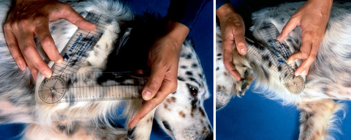
Extend and flex the carpus. Maximum extension of the carpus should be about 180° to 190°; maximum flexion should be 45°.
- A decreased range of motion may indicate degenerative joint disease. Note any crepitation.
- Extend and stress the carpus in the medial to lateral plane to determine if there is joint instability. Also observe the dog while standing to determine if hyperextension is present.

Radius:
Palpate the radius for instability (fracture), swelling (fracture, tumor), and/or pain response to deep bone palpation (panosteitis).
Elbow:
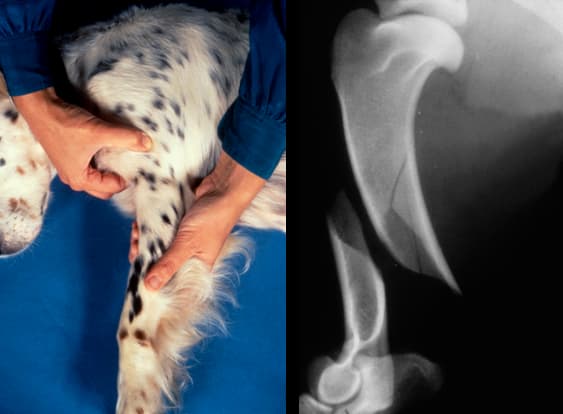
a. )Palpate the elbow for fluctuant swelling in the space between the lateral condyle and olecranon, and over the medial coronoid process. This indicates joint effusion which can result from several elbow diseases.
Joint effusion may be more easily detected when the animal is standing. Firm, generalized swelling of the elbow often indicates degenerative joint disease.
(b. and c.) Flex and extend the elbow. Normal extension and flexion is about 165° and 40° to 50°, respectively. The carpus should almost touch the shoulder when the elbow is flexed. While the elbow is in extension check the integrity of the collateral ligaments by applying medial and lateral force to the radius and ulna.

Decreased range of motion caused by incomplete flexion of the elbow usually suggests degenerative joint disease.
Degenerative joint disease of the elbow is usually secondary to a fragmented coronoid process, an ununited anconeal process, or osteochondritis dissecans.
Humerus:
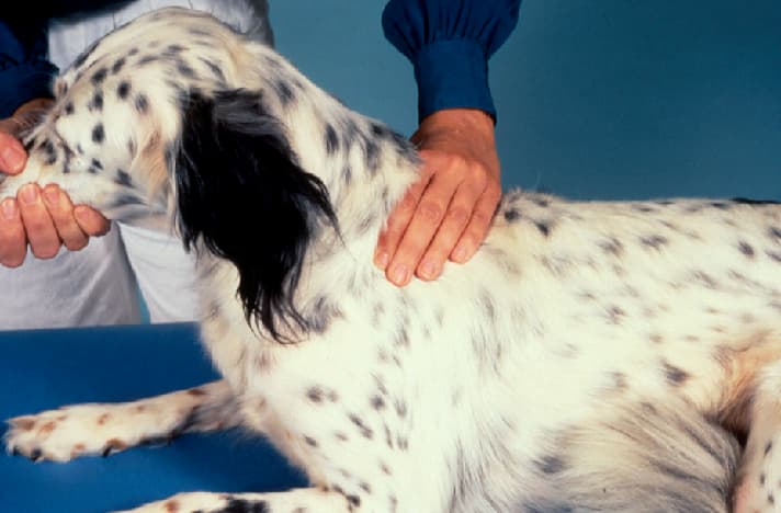
Palpate the humerus for instability (fracture), swelling (fracture, tumor) and pain response to deep palpation (panosteitis). Palpate the bone in areas where it is not covered by muscles to differentiate panosteitis from muscular pain.
Shoulder:
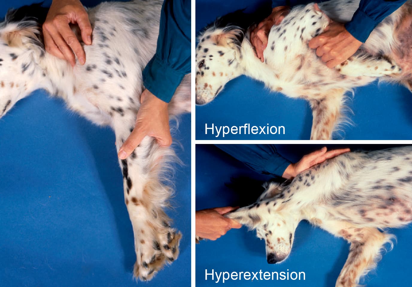
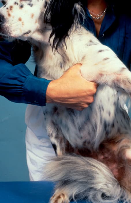
Shoulder Swelling from joint effusion is difficult to detect in the shoulder because of the overlying muscles. Move the shoulder through a range of motion, including hyperextension and hyperflexion while stabilizing the scapula.
Scapula:
Palpate the scapula for instability (fracture) and swelling (fracture, tumor). Palpate the muscle over the scapula and compare it to the opposite side to determine atrophy secondary to disuse or nerve injury. Probe the axillary area for swellings and observe for signs of pain. This can be indicative of a nerve root tumor.