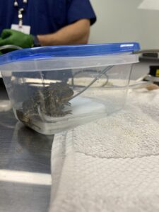
This adult male American toad (Bufo americanus) presented to the Wildlife Medical Clinic in November with a left hind leg wound. While the injury was notably severe, our exam showed the toad was using the leg to bear weight and had appropriate reflexes, indicating that the nerves were still functional. Despite this, his leg wound turned out to be a fracture with bone exposure. His wounds were treated topically and pain medication administered while we devised a longer term plan for his care. Amphibians are uncommon WMC patients, and we had to do some research to see how we could help this toad!
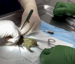
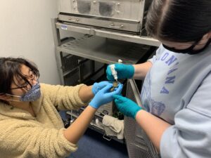
After discussing the case with one of our veterinarians, we pursued radiographs to see how healthy the left hind leg was internally. This would help us determine if we could potentially correct the fracture or amputate part of the leg, removing the diseased part and maintaining as much of the leg as possible for mobility. We had to sedate the toad to get the best quality radiographs. Since amphibians can quickly absorb medications across their skin, we used a special anesthetic called tricaine methanesulfonate (more commonly called MS-222). This drug is dissolved in water and absorbed across the gills or skin of fish and amphibians, respectively, as an anesthetic.
The toad sat in the solution and absorbed the anesthetic through his skin while we monitored him for signs of sedation, including slower breathing, less muscle tone, and loss of reflexes. When he was adequately sedate, we performed radiographs. Those images showed us the calcaneum (an ankle bone) was luxated and protruding from the skin while some of the outer metatarsal bones (toes) were no longer present. In order to include as much healthy skin and bone as possible, we decided to amputate the leg at the mid-tibiofibula (mid-shin) while the toad was still under anesthesia. During the surgery, the patient was monitored using a Doppler probe to listen to the heart. The amputation went well and left a good amount of the leg as a mobility aid. In addition to pain medications, this toad was treated with antibiotics as he healed.
In the days after his surgery, this toad developed a cloudy right eye. It was initially evaluated for a possible ulcer and, having none, treated symptomatically with eye drops to help draw out the inflammation our ophthalmic (eye) exam found. Despite our treatments, his eye continued to worsen, eventually developing a pyramided appearance. Examination of the eye by a veterinary ophthalmologist determined this frog had bullous keratopathy a condition in which the cornea (outer surface of the eye) are covered in bullae or blisters. To visualize these bullae, a microscopic examination of the eye with a special instrument called a slit lamp was necessary. Staining the eye showed that none of the blisters had burst and caused an ulcer yet, and examination of the other structures of the eye showed no disease deep inside the eye, only the bullae on the cornea. As he had failed to improve with our topical treatments, the next option for this toad was to perform a tarsorrhaphy. In this procedure, the eyelid is temporarily sealed over the eye to apply a constant, light pressure on the bullae to reduce them while simultaneously protecting the eye. Since toads do not have mobile eyelids, this procedure could be done with the false third eyelid which is a thin membrane that closes upwards over the cornea to protect and lubricate the eye. The procedure required another anesthesia and was a success!
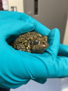
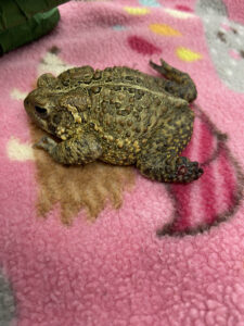
While this toad threw us a curve ball or two during his healing, we are happy to report he is well on his road to recovery. His amputation site has healed completely, and he is moving around well. Additionally, his eye has completely healed and fully functional again! This toad will spend several more months with us as we wait for the weather to warm up enough to allow for his release. This case has presented a wonderful learning opportunity for all of the Wildlife Medical Clinic students to learn about amphibian medicine, orthopedic surgery, anesthesiology, and ophthalmology. We are grateful to the finder who brought him to us, surely saving this toad’s life and facilitating a great learning experience in the process!
Written By: Billy Wright Class of 2024