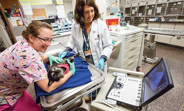
Above: Dr. Caroline Tonozzi, right, performs point-of-care ultrasound on a dog.
In the emergency and critical care service at the Veterinary Teaching Hospital, we work hard to train our students and house officers to triage and treat critically ill patients effectively.
A triage exam involves collecting patient information that could significantly influence decisions about emergency treatment. In addition to the hands-on physical exam, we use modalities called point-of-care testing, such as the Nova Biomedical portable blood gas analyzer and point-of-care ultrasound (POCUS), to assist us on making lifesaving decisions for our patients.
POCUS is versatile and easy to perform tableside for our patients. We use it with nearly every patient, just as we would use an ECG or blood pressure machine. A point-of-care ultrasound is done cageside or tableside during patient resuscitation in order to gain patient information beyond what was noted on the triage exam.
Patients presenting to the hospital with a history of collapse, trauma, respiratory distress, or abdominal pain and patients near cardiopulmonary arrest benefit from POCUS. It may also confirm our suspicion for findings such as the presence or absence of free fluid in the pericardial, pleural, or peritoneal spaces or a pneumothorax.
POCUS may also identify findings that assist clinicians with the pursuit of a diagnosis in an unstable patient. However, we must emphasize that this modality does not replace more conventional imaging modalities such as radiography or traditional ultrasonography of the abdomen or thorax.
Specific POCUS of the thorax and abdomen, when used in combination, complement each other, and we use them together very commonly in our patients. Focused Assessment with Sonography for Triage, or FAST, allows a clinician to very quickly assess a thorax (TFAST) or abdomen (AFAST) for the presence or absence of free fluid.
Vet BLUE® (for “bedside lung ultrasound examination) allows a clinician to scan the lungs, in addition to the pleural space, when performing a TFAST to look for pulmonary parenchymal changes. For a patient presenting in respiratory distress with a heart murmur, the clinician may perform a La:Ao (left atrial-to-aortic) ratio assessment to determine if furosemide should be given. Noting the caudal vena cava distension or collapse may assist a clinician in deciding whether a patient is normovolemic or requires additional fluid therapy.
Ultimately this information done tableside benefits the patient and the client. We can quickly provide estimates of cost and set reasonable expectations for owners as they navigate the difficult road of critical illness.
The emergency department isn’t the only place where POCUS is useful. Critically ill patients in our ICU may require US-guided venous access when placing a jugular catheter or to measure urinary bladder size. Patients that acutely worsen may benefit from a FAST scan to determine if the accumulation of free fluid is the cause for the acute change, especially in the postoperative GI patient. A clinician may utilize POCUS to try to figure out why a patient is declining in the face of aggressive resuscitation.
Point of care ultrasound is easy to perform cageside and provides a wealth of information regarding patient status.
Reach the ER/ICU at 217-333-5311 to alert us when you are referring a case.
By Caroline Tonozzi, DVM, DACVECC