The soft tissue surgery team at the Veterinary Teaching Hospital is excited to introduce our new minimally invasive surgery service. After training in Canada with a fellow of the Veterinary Endoscopy Society in advanced laparoscopic and thoracoscopic surgeries, I joined the Illinois faculty last fall and will head the minimally invasive surgery service.
With the latest equipment and our brand-new surgery wing, we are now able to offer cutting-edge minimally invasive procedures such as laparoscopic adrenalectomy, thoracoscopic ligamentum arteriosum transection for PRAA, and thoracic duct ligation for chylothorax.
While we are proud to offer a wide range of procedures, we have highlighted a few to illustrate the advantages of a minimally invasive approach.
Laparoscopic Cholecystectomy
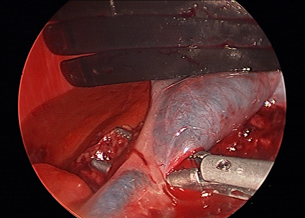
Gallbladder mucoceles are the most common extrahepatic surgical disease in our canine companions. The clinical signs may vary from an incidental finding to hypotensive shock. Surgical intervention is preferred in earlier uncomplicated cases. Laparoscopic cholecystectomy is ideal in these patients to minimize morbidity and hospitalization times.
In the intraoperative image of a laparoscopic cholecystectomy above, the gall bladder is retracted dorsally with a fan retractor while the cystic duct is being dissected with articulating forceps.
Thoracoscopic Lung Lobectomy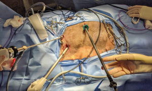
While primary lung neoplasia is much less common than metastatic disease, the prognosis with surgical removal can be good. A traditional lung lobectomy involves a large intercostal incision with spreading of the ribs, which can be extremely painful and lead to prolonged recovery times for these older patients. Thoracoscopic lung lobectomy requires only three to four 1-cm incisions, significantly reducing post-operative while improving visualization to excise the primary tumor with clean margins, and any metastatic lymph nodes.
The intraoperative image at right shows the port placement for a left caudal lung lobectomy.
This patient had four 0.5- to 1-cm incisions to facilitate placement of the thoracoscope, stapling device, and instruments. She recovered without complication and was discharged the following day.
Total Laparoscopic Gastropexy
While laparoscopic-assisted gastropexies are commonly performed as a prophylactic measure against gastric dilatation and volvulus, total laparoscopic gastropexy has been shown to have less impact on post-operative activity. A laparoscopic-assisted gastropexy involves a 6-cm incision made in the right para-costal region; the gastropexy is performed by suturing the stomach serosa and muscularis to the transversus abdominal muscle extracorporeally (outside of the abdomen).
By contrast, in total laparoscopic gastropexy, three 0.5-cm incisions are made on ventral midline to facilitate placement of needle drivers and a laparoscope. The gastropexy is performed intracorporeally (completely within the abdomen) with barbed suture.
The video below is a three-minute video of a total laparoscopic gastropexy. You can watch as the suturing is performed intracorporeally. The patient is left with only three small external incisions.
If you are interested in referring a patient or learning more about minimally invasive procedures, please call us at 217-333- 6808 or email me at jscott19@illinois.edu.
By Jacqui Scott, DVSc, DACVS (Small Animal), MANZCVSc